Saizonou MA, Kitazawa H, Kanahashi T, Yamada S, Takakuwa T, Epithelial development of the urinary collecting system in the human embryo, PLOS ONE 19(4): e0301778. https://doi.org/10.1371/journal.pone.0301778
Abstract
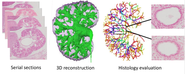
The urinary collecting system (UCS) consists of organized ducts that collect urine from the nephrons and transport it to the ureter and bladder. Understanding the histogenesis of the UCS is critical. Thirty human embryos between the Carnegie stages (CS) 18 and 23 were selected from the Congenital Anomaly Research Center, Kyoto, Japan. Epithelia of the UCS, ureter, and bladder of each sample were randomly selected. Histological findings of the epithelia were analyzed according to the following criteria: type of epithelium, presence or absence of glycogen, percentage of migrated nuclei, percentage of cells in mitosis, and the surrounding mesenchyme. A thickened epithelium lining a narrow luminal cavity was observed in the pre-expanded pelvic specimens at CS18-CS23. At CS23, after pelvic expansion, the UCS showed a thin epithelium with a large luminal cavity mainly located on the early branches, whereas the epithelium covering the subsequent branches had medium thickness. Histological characteristics differed depending on the UCS part and sample stage. The degree of differentiation was evaluated, revealing that in CS18-CS23 pre-expanded pelvis specimens, the undifferentiated epithelium was found in the zeroth to third/fifth generation, whereas at CS23, after pelvic expansion, a differentiated epithelium covered the UCS zeroth to seventh generation. In a comparison of the urothelial epithelium between the UCS, ureter, and bladder, we found that urinary tract differentiation may be initiated in the bladder, followed by the ureter, UCS zeroth to seventh generations, and finally, UCS eighth to end generations. An understanding of the histogenesis of embryonic stage UCS can aid in the clinical management of congenital urinary tract defects and other diseases.
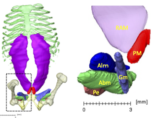
The pyramidalis muscle (PM) is a paired small triangular muscle of the anterior abdominal wall, the physiological significance of which remains unclear. Recent studies have failed to detect this muscle during the embryonic period. Hence, the present study aimed to determine when PM emerged and reveal its features using high-resolution magnetic resonance imaging. Fourteen embryos between Carnegie stage (CS)18 and CS23 and 59 fetuses (crown-rump length: 39.5–185.0 mm) were selected for this study. The PM was first detected in one of the three samples at CS20. It was detected in five of the seven samples (71.4%) between CS21 and CS23. Forty-eight samples (81.4%) at early fetal period had PMs on both the right and left sides, and three (5.1%) had that only on the right side. Eight samples (13.6%) had no PMs. No side-differences or sexual dimorphisms were detected. The PM length was larger than the width in most samples, although the length/width ratio varied among the samples. The PM/rectus abdominis muscle length and PM/umbilicus-pubic symphysis length ratios were almost constant, irrespective of the crown-rump length. The PM is located ventrally inferior to the rectus abdominis and closer to the medial muscle groups of the lower limb than the rectus abdominis. The present study demonstrated that PM formation occurred in the late embryonic period, and that the frequency, side differences, sex dimorphism, and spatial position of the PM in the early fetal period were similar to those in adults.
Fujii S, Muranaka T, Matsubayashi J, Yamada S, Yoneyama A, Takakuwa T. Bronchial tree of the human embryo: Examination based on a mammalian model. J Anatomy 2024, 244, 159-169 http://doi.org/10.1111/joa.13946 .
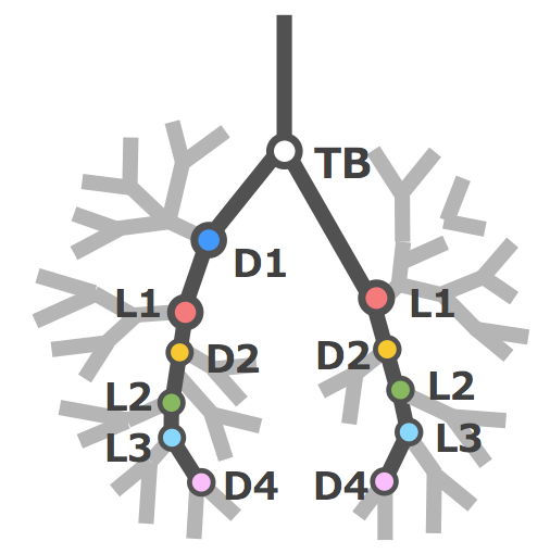
The symmetry of right and left bronchi, proposed in a previous comparative anatomical study as the basic model of the mammalian bronchial tree, was examined to determine if it applied to the embryonic human bronchial tree. Imaging data of 41 human embryo specimens at Carnegie stage (CS) 16–23 (equivalent to 6–8 weeks after fertilization) belonging to the Kyoto collection were obtained using phase-contrast X-ray computed tomography. Three-dimensional bronchial trees were then reconstructed from these images. Bronchi branching from both main bronchi were labeled as dorsal, ventral, medial, or lateral systems based on the branching position with numbering starting cranially. The length from the tracheal bifurcation to the branching point of the labeled bronchus was measured, and the right-to-left ratio of the same labeled bronchus in both lungs was calculated. In both lungs, the human embryonic bronchial tree showed symmetry with an alternating pattern of dorsal and lateral systems up to segmental bronchus B9 as the basic shape, with a more peripheral variation. This pattern is similar to that described in adult human lungs. Bronchial length increased with the CS in all labeled bronchi, whereas the right-to-left ratio was constant at approximately 1.0. The data demonstrated that the prototype of the human adult bronchial branching structure is formed and maintained in the embryonic stage. The morphology and branching position of all lobar bronchi and B6, B8, B9, and the subsegmental bronchus of B10 may be genetically determined. On the other hand, no common structures between individual embryos were found in the peripheral branches after the subsegmental bronchus of B10, suggesting that branch formation in this region is influenced more by environmental factors than genetic factors.
Fukui N, Toru KanahashiT, MatsubayashiJ, ImaiH, YoneyamaA, OtaniH, YamadaS, Takakuwa T. Morphogenesis of the pulmonary vein and left atrial appendage in human embryos and early fetuses. J Anatomy 2024, 244, 142-158, in press, https://doi.org/10.1111/joa.13941
Abstract
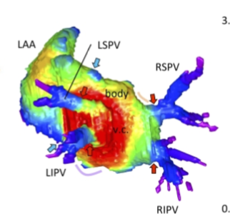
The left atrium wall has several origins, including the body, appendage, septum, atrial–ventricular canal, posterior wall, and venous component. Here, we describe the morphogenesis of left atrium based on high-resolution imaging (phase-contrast X-ray computed tomography and magnetic resonance imaging). Twenty-three human embryos and 19 fetuses were selected for this study. Three-dimensional cardiac images were reconstructed, and the pulmonary veins and left atrium, including the left atrial appendage, were evaluated morphologically and quantitatively. The positions of the pericardial reflections were used as landmarks for the border of the pericardial cavity. The common pulmonary vein was observed in three specimens at Carnegie stage 17–18. The pericardium was detected at the four pulmonary veins (left superior, left inferior, right superior, and right inferior pulmonary veins) at one specimen at Carnegie stage 18 and all larger specimens, except the four samples. Our results suggest that the position of the pericardial reflections was determined at two pulmonary veins (right and left pulmonary vein) and four pulmonary veins almost simultaneously when the dorsal mesocardial connection between the embryo and heart regressed. The magnetic resonance images and reconstructed heart cavity images confirmed that the left atrium folds were present at the junction between the body and venous component. Three-dimensional reconstruction showed that the four pulmonary veins entered the dorsal left atrium tangentially from the lateral to the medial direction. More specifically, the right pulmonary veins entered at a greater angle than the left pulmonary veins. The distance between the superior and inferior pulmonary veins was shorter than that between the left and right pulmonary veins. Three-dimensional reconstruction showed that the venous component increased proportionally with growth. No noticeable differences to discriminate between the right and left parts of the venous component emerged, while the junction between the venous component and body gradually became inconspicuous but was still recognizable by the end of the observed early fetal period. The left superior pulmonary vein had the smallest cross-sectional area and most flattened shape, whereas the other three were similar in area and shape. The left atrial appendage had a large volume in the center and extended to the periphery as a lobe-like structure. The left atrial appendage orifice increased in the area and tended to become flatter with growth. The whole left atrium volume^(1/3) increased almost proportionally with growth, parallel to the whole heart volume. This study provided a three-dimensional and quantitative description of the developmental process of left atrium, comprising the venous component and left atrial appendage formation, from the late embryonic to the early fetal stages.
60. Takakuwa T, Saizonou MA, Fujii S, Kumano Y, Ishikawa A, Aoyama T, Imai H, Yamada S, Kanahashi T. Femoral posture during embryonic and early fetal development: An analysis using landmarks on the cartilaginous skeletons of ex vivo human specimens. PLOS one, 2023, 18(5): e0285190. https://doi.org/10.1371/journal.pone.0285190.
Abstract
The pre-axial border medially moves between the fetal and early postnatal periods, and the foot sole can be placed on the ground. Nonetheless, the precise timeline when this posture is achieved remains poorly understood. The hip joint is the most freely movable joint in the lower limbs and largely determines the lower-limb posture. The present study aimed to establish a timeline of lower-limb development using a precise measurement of femoral posture. Magnetic resonance images of 157 human embryonic samples (Carnegie stages [CS] 19–23) and 18 fetal samples (crown rump length: 37.2–225 mm) from the Kyoto Collection were obtained. Three-dimensional coordinates of eight selected landmarks in the lower limbs and pelvis were used to calculate the femoral posture. Hip flexion was approximately 14° at CS19 and gradually increased to approximately 65° at CS23; the flexion angle ranged from 90° to 120° during the fetal period. Hip joint abduction was approximately 78° at CS19 and gradually decreased to approximately 27° at CS23; the average angle was approximately 13° during the fetal period. Lateral rotation was greater than 90° at CS19 and CS21 and decreased to approximately 65° at CS23; the average angle was approximately 43° during the fetal period. During the embryonic period, three posture parameters (namely, flexion, abduction, and lateral rotation of the hip) were linearly correlated with each other, suggesting that the femoral posture at each stage was three-dimensionally constant and exhibited gradual and smooth change according to growth. During the fetal period, these parameters varied among individuals, with no obvious trend. Our study has merits in that lengths and angles were measured on anatomical landmarks of the skeletal system. Our obtained data may contribute to understanding development from anatomical aspects and provide valuable insights for clinical application.
59. Matsunari C, Kanahashi T, Otani H, Imai H, Yamada S, Okada T, Takakuwa T. Tentorium cerebelli formation during human embryonic and early fetal development. Anat Rec (Hoboken) 2023, 306(3), 515-526
Abstract
The morphologies of the fetal tentorium cerebelli (TC) and brain influence each other during development. This study aimed to analyze and more comprehensively understand the three-dimensional morphogenesis of the TC and fetal brain. We examined magnetic resonance imaging from 64 embryonic and fetal specimens (crown-rump length range, 9.2–225 mm). During the embryonic period, the lateral folds of the TC elongated to traverse the middle part of the midbrain. The TC and falx cerebri appeared separated, and no invaginations at the parieto-occipital region were observed. In the early fetal period, the cerebrum covered approximately half of the midbrain. The separation of the dural limiting layer at the parieto-occipital region widened from the posterior cerebrum to the cranial cerebellum. The lateral folds of the TC were spread between its tip, continuous with the falx cerebri, and its base plane, located between the midbrain and rostral hindbrain. Differences in the TC components’ growth directions gradually diminished as the cerebrum covered the midbrain. We observed rotation of the TC at its median section according to its growth, which ceased in the middle fetal period. The brainstem and cerebellum extended inferiorly via differential growth, with the cerebrum covering them superiorly. The morphology of the TC curved to conform to the cerebellar and cerebral surfaces. Our present study suggests that factors affecting TC morphology differ between the early and middle fetal periods. Present data provided a more comprehensive view of TC formation according to developmental stage.
Kanahashi T, Imai H, Otani H, Yamada S, Yoneyama A, Takakuwa T. Three-dimensional morphogenesis of the human diaphragm during the late embryonic and early fetal period: Analysis using T1-weighted and diffusion tensor imaging. J Anat. 2023, 242, 174-190, DOI: 10.1111/joa.13760
Abstract
表紙に採用されました
A precise understanding of human diaphragm development is essential in fetal medicine. To our knowledge, no previous study has attempted a three-dimensional (3-D) analysis and evaluation of diaphragmatic morphogenesis and development from the embryonic to the early fetal period. This study aimed to evaluate the morphogenesis and fibrous architecture of the developing human diaphragm during the late embryonic and early fetal periods. Fifty-seven human embryos and fetuses (crown-rump length [CRL] = 8–88 mm) preserved at the Congenital Anomaly Research Center of Kyoto University and Shimane University were analyzed. 3-D morphogenesis and fiber orientation of the diaphragm were assessed using phase-contrast X-ray computed tomography, T1-weighted magnetic resonance imaging (T1W MRI), and diffusion tensor imaging (DTI). T1W MR images and DTI scans were obtained using a 7-T MR system. The diaphragm was completely closed at Carnegie stage (CS) 20 and gradually developed a dome-like shape. The diaphragm was already in contact with the heart and liver ventrally in the earliest CS16 specimen observed, and the adrenal glands dorsally at CS19 or later. In the fetal period, the diaphragm contacted the gastric fundus in samples with a CRL ≥41 mm, and the spleen in samples with a CRL ≥70 mm. The relative position of the diaphragm with reference to the vertebrae changed rapidly from CS16 to CS19. The most cranial point of the diaphragm was located between the 4th and 8th thoracic vertebrae, regardless of fetal growth, in samples with a CRL ≥16 mm. Diaphragmatic thickness was nearly uniform (0.15–0.2 mm) across samples with a CRL of 8 mm to 41 mm. The sternal, costal, lumbar parts, and the area surrounding the esophageal hiatus thickened with growth in samples with a CRL ≥46 mm. The thickness at the center of the diaphragm and the left and right hemidiaphragmatic domes did not increase with growth. Tractography showed that the fiber orientation of the sternal, costal, and lumbar parts became more distinct as growth progressed in CS19 or later. All fibers in the costal and lumbar regions ran toward the left and right hemidiaphragmatic domes, except for those running to the caval opening and esophageal hiatus. The fiber orientation patterns from the right and left crura surrounding the esophageal hiatus were classified into three types. Distinct fiber directions between the costal and sternal, and between the costal and lumbar diaphragmatic parts were observable in samples with a CRL ≥46 mm. Anterior costal and sternal fibers ran toward the center. Fiber tracts around the center and the left and right hemidiaphragmatic domes; between the costal and lumbar orientations; and between the costal and sternal orientations showed a tendency for decreasing fractional anisotropy values with fetal growth, and showed less density than other areas. In conclusion, we used 3-D thickness assessment and DTI tractography to identify qualitative changes in the muscular and tendonous regions of the diaphragm during the embryonic and early fetal periods. This study provides information on normal human diaphragm development for the progression of fetal medicine and furthering the understanding of congenital anomalies.
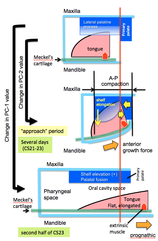
Nohara A, Owaki N, Matsubayashi J, Katsube M, Imai H, Yoneyama A, Yamada S, Kanahashi T, Takakuwa T. Morphometric analysis of secondary palate development in human embryos. J Anatomy, 2022, 241(6), 1287-1302, 2022, DOI:10.1111/joa.13745
Abstract
Rapid shelf elevation and contact of the secondary palate and fusion reportedly occur due to a growth-related equilibrium change in the structures within the oro-nasal cavity. This study aimed to quantitatively evaluate complex three-dimensional morphological changes and their effects on rapid movements, such as shelf elevation and contact, and fusion. Morphological changes during secondary palate formation were analyzed using high-resolution digitalized imaging data (phase-contrast X-ray computed tomography and magnetic resonance images) obtained from 22 human embryonic and fetal samples. The three-dimensional images of the oro-nasal structures, including the maxilla, palate, pterygoid hamulus, tongue, Meckel’s cartilage, nasal cavity, pharyngeal cavity, and nasal septum, were reconstructed manually.
palatal shelves were not elevated in all the samples at Carnegie stage (CS)21 and CS22 and in three samples at CS23. In contrast, the palatal shelves were elevated but not in contact in one sample at CS23. Further, the palatal shelves were elevated and fused in the remaining four samples at CS23 and all three samples from the early fetal period. For each sample, 70 landmarks were subjected to Procrustes and principal component (PC) analysis. PC-1 accounted for 67.4% of the extracted gross changes before and after shelf elevations. Notably, the PC-1 values of the negative and positive value groups differed significantly. The PC-2 value changed during the phases in which the change in the PC-1 value was unnaturally slow and stopped at CS22 and the first half of CS23. This period, defined as the “approach period”, corresponds to the time before dynamic changes occur as the palatal shelves elevate, the tongue and mandibular tip change their position and shape, and secondary palatal shelves contact and fuse. During the “approach period”, measurements of PC-2 changes showed that structures on the mandible (Meckel’s cartilage and tongue) and maxilla (palate and nasal cavity) did not change positions, albeit both groups of structures appeared to be compressed anterior-posteriorly. However, during and after shelf elevation, measurements of PC-1 changes showed significant changes between maxillary and mandibular structures, particularly positioning of the shelves above the tongue and protrusion of the tongue and mandible. These results suggest an active role for Meckel’s cartilage growth in repositioning the tongue to facilitate shelf elevation. The present data representing three distinct phases of secondary palate closure in humans can advance the understanding of morphological growth changes occurring before and after the horizontal positioning of palatal shelves and their fusion to close the secondary palate in humans successfully.