Ohga A, Sakamoto R, Yamada S, Takakuwa T, Vesicular swelling in the cervical region with lymph sac formation in human embryos. Congenit Anom, 2020, 60, 62-67: 10.1111/cga.12339.
Abstract
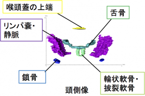
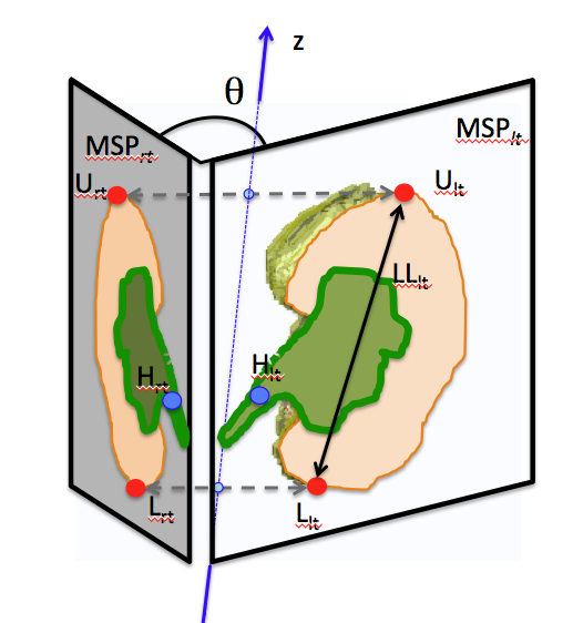
Vesicular swelling in the cervical region (VSC) is occasionally observed among human embryos around Carnegie stage (CS) 21. However, its mechanism and significance in fetal development are unclear. The present study aimed to analyze the relation of development of VSC with jugular lymph sac (JLS) formation. Serial histological sections that were digitalized from 14 embryos at CS20 and CS21 stored at the Kyoto Collection were used for the analysis. Subcutaneous edema and enlargement of the subarachnoid space were found to cause VSC. No obvious abnormalities in cranial regions that may be related to the VSC were detected on histological sections. Three-dimensional reconstructions revealed the following: (a) the JLS was located bilaterally at the levels between the first and fourth cervical vertebrae; (b) the JLS was pyramidal in shape; and (c) no severe deformity and/or malformation was found in all samples. The JLS was not connected to the subcutaneous tissue and subarachnoid space in all samples. The mean volume of the JLS increased nine-times from CS20 (0.02 mm3 in VSC [−] group) to CS21 (0.18 mm3 in VSC [−] group). The mean volume of the JLS was comparable between the VSC [−] and VSC (+) groups at both CS20 and CS21. A moderate correlation was observed between VSCd and the mean volume of the JLS in both groups at CS20 (R2 = 0.75) and CS21 (R2 = 0.56). In conclusion, the dynamics of the lymphatic system at the cervical region may contribute to VSC observed around CS21.
40. Matsubayashi J, Okuno K, Fuji S, Ishizu K, Yamada S, Yoneyama A, Takakuwa T. Human embryonic ribs all progress through common morphological forms irrespective of their position on the axis, Dev Dyn 2019, 248, 1257-1263, doi: 10.1002/dvdy.107
parsimonious model
Abstract
Background
We aimed to analyze the morphogenesis of all ribs from 1st to 12th rib pairs plus vertebrae to compare their differences and features according to the position along the cranial-caudal axis during the human embryonic period.
Results
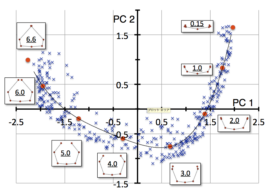
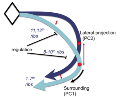
Rib pair formation was analyzed using high-resolution digitalized imaging data (n = 29) between Carnegie stage (CS) 18 and CS23 (corresponding to ED13-14 in mouse; HH29-35 in chick). A total of 348 rib pairs, from 1st to 12th rib pairs of each sample were subjected to Procrustes and principal component (PC) analyses. PC1 and PC2 accounted for 76.3% and 16.4% (total 92.7%) of the total variance, respectively, indicating that two components mainly accounted for the change in shape. The distribution of PC1 and PC2 values for each rib showed a “fishhook-like shape” upon fitting to a quartic equation. PC1 and PC2 value position for each rib pair moved along the fitted curve according to the development. Thus, the change in PC1 and PC2 could be expressed by a single parameter using a fitted curve as a linear scale for shape.
Conclusion
Human embryonic ribs all progress through common morphological forms irrespective of their position on the axis.
Okuno K, Ishizu K, Matsubayashi J, Fujii S, Sakamoto R, Ishikawa A, Yamada S, Yoneyama A, Takakuwa T. Rib cage morphogenesis in the human embryo: A detailed three-dimensional analysis. Anat Rec 2019, 302, 2211-2223, doi: 10.1002/ar.24226
ABSTRACT
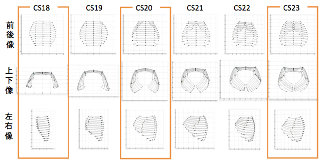
Formation of the skeletal structure in the human embryo has important consequences in terms of support, protection, and function of organs and other systems. We aimed to describe the formation of the rib cage during the embryonic period, in order to detect prominent features and identify the possible factors affecting rib cage morphology. We employed high-resolution digitized imaging data (n = 34) obtained in human embryos with Carnegie stage (CS) between 17 and 23. The rib cage became detectable as cartilage formation at CS17, expanding outward from the dorsal side of the chest-abdominal region. Ribs elongated progressively to surround the chest, differentiating into the upper and lower rib cage regions by CS20. The ends of corresponding ribs in the upper region elongated toward each other, leading to their joining and sternum formation between CS21 and CS23, while the lower region of the rib cage remained widely open. The rib cage area with the largest width shifted from the 5th rib pair at CS17 to the 9th pair at CS23. The depth of the rib cage was similar across the upper region at CS17, with the major portion remaining in the middle part after CS20. The heart was located beneath the rib pairs providing the largest depth, while the liver was located beneath the rib pairs providing the largest width. Formation of the sternum, development of spinal kyphosis, and organization of larger internal organs within the thoracic and abdominal cavity are possible factors affecting rib cage morphology. Anat Rec, 302:2211–2223, 2019.
37. Ishiyama H, Ishikawa A, Imai H, Matsuda T, Yoneyama A, Yamada S, Takakuwa T. Spatial relationship between the metanephros and adjacent organs according to the Carnegie stage of development. Anat Rec 2019. 302, 1887-2104. DOI: 10.1002/ar.24103
ABSTRACT
The morphological changes in the metanephros and its spatial relationship to the adjacent organs was evaluated based on the Carnegie stages (CSs) from 14 through 23. The imaging modalities used included magnetic resonance imaging (N = 4), phase-contrast X-ray computed tomography (N = 11), and serial histological sections (N = 40), supplemented by three-dimensional image reconstruction. The orientation of the hilus of the metanephros changed significantly between CS17 (34.4 ± 13.7 degrees) and 18 (122.3 ± 38.1 degrees), with an increase in the number of branches of the urinary collecting system, from 1.61 ± 0.42 at CS17 to 3.20 ± 0.35 at CS18. This increase in the number of branches influenced the growth of the metanephros and the orientation of its hilus. The right and left metanephroses were in proximity throughout the embryonic period. The local maximum interpole distances were observed at CS18 (0.87 ± 0.11 mm for the upper and 0.50 ± 0.25 mm for the lower pole). Mesenchymal tissue was observed between the metanephros and iliac arteries, as well as between the right and left metanephros. Throughout development, the position of the lower pole of the metanephros remained adjacent to the aortic bifurcation. The position of the upper pole, referenced with respect to the aortic bifurcation, increased by >2.0 mm, reflecting the longitudinal growth of the metanephros. Our findings provide a detailed description of the morphogenesis of the metanephros and of its hilus, which might contribute to our understanding of congenital malformations and malpositions of the kidneys.
Kanahashi T, Yamada S, Yoneyama A, Takakuwa T. Relationship Between Physiological Umbilical Herniation and Liver Morphogenesis During the Human Embryonic Period: A Morphological and Morphometric Study. Anat Rec 2019, 302, 1968-1976. doi: 10.1002/ar.24149.
肝臓無形成でも生理的臍帯ヘルニアがみられる
ABSTRACT
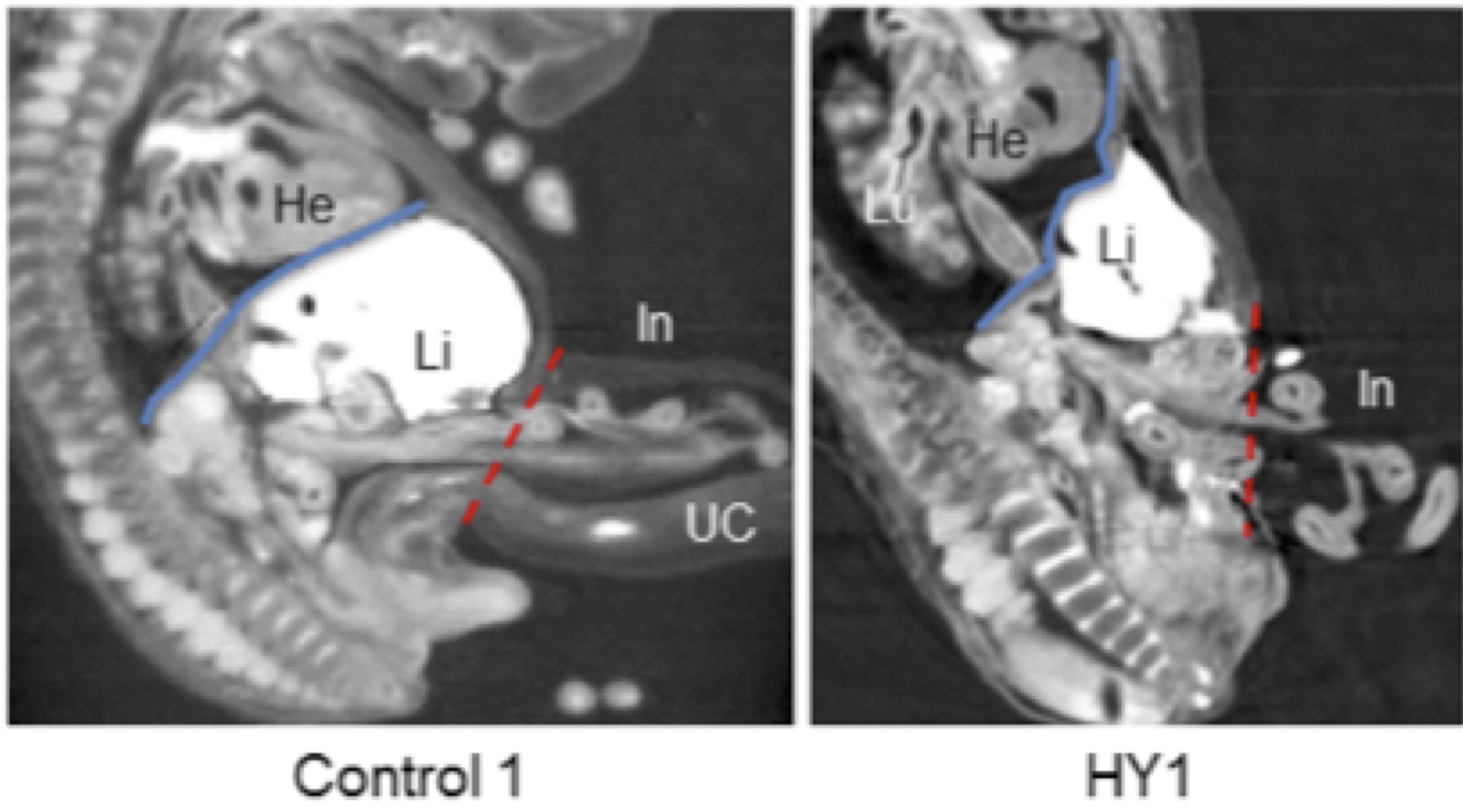
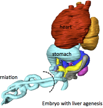
It is widely hypothesized that physiological umbilical herniation (PUH) in humans occurs, because the liver occupies a large space in the abdominal cavity, which pushes the intestine into the extraembryonic coelom during the embryonic period. We have recently shown the presence of the intestinal loop in the extraembryonic coelom in embryos with liver malformation. Here, we analyzed the relationship between the liver and the PUH at Carnegie stage 21 of four embryos with liver malformation, including two with hypogenesis (HY1, HY2) and two with agenesis (AG1, AG2), using phase-contrast X-ray computed tomography and compared them with two control embryos. The intestinal loop morphology in the malformed embryos differed from that in the control embryos, except in HY1. The length of the digestive tract in the extraembryonic coelom of the embryos with liver malformation was similar to or longer than that of the controls. The rate of intestinal loop lengthening in the extraembryonic coelom compared with that of the total digestive tract in all embryos with liver malformation was similar to or higher than that of the controls. The estimated total abdominal cavity volume in the embryos with liver malformation was considerably smaller than that of the controls, while the intestinal volume was similar. The cardia and proximal portion of the pancreas connecting to the duodenum were located at almost identical positions in all the embryos, whereas other parts of the upper digestive tract deviated in the embryos with abnormal livers. Thus, our results provided evidence that PUH occurred independently of liver volume.
Suzuki Y, Matsubayashi J, Ji X, Yamada S, Yoneyama A, Imai H, Matsuda T, Aoyama T, Takakuwa T Morphogenesis of the femur at different stages of normal human development, PLoS ONE, 14(8): e0221569. https://doi.org/10.1371/journal. pone.0221569
Abstract
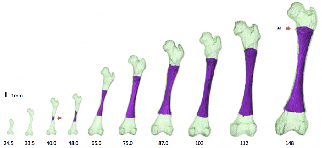
The present study aimed to better characterize the morphogenesis of the femur from the embryonic to the early fetal periods. Sixty-two human fetal specimens (crown–rump length [CRL] range: 11.4–185 mm) from the Kyoto Collection were used for this study. The morphogenesis and internal differentiation process of the femur were analyzed in 3D using phase-contrast X-ray computed tomography and magnetic resonance imaging. The cartilaginous femur was first observed at Carnegie stage 18. Major anatomical landmarks were formed prior to the initiation of ossification at the center of the diaphysis (CRL, 40 mm), as described by Bardeen. The region with very high signal intensity (phase 5 according to Streeter’s classification; i.e., area described as cartilage disintegration) emerged at the center of the diaphysis, which split the region with slightly low signal intensity (phase 4; i.e., cartilage cells of maximum size) in fetuses with a CRL of 40.0 mm. The phase 4 and phase 5 regions became confined to the metaphysis, which might become the epiphyseal cartilage plate. Femur length and ossified shaft length (OSL) showed a strong positive correlation with CRL. The OSL-to-femur length ratio rapidly increased in fetuses with CRL between 40 and 75 mm, which became moderately increased in fetuses with a CRL of ≥75 mm. Cartilage canal invasion occurred earlier at the proximal epiphysis (CRL, 62 mm) than at the distal epiphysis (CRL, 75 mm). Morphometry and Procrustes analysis indicated that changes in the femur shape after ossification were limited, which were mainly detected at the time of initial ossification and shortly after that. In contrast, femoral neck anteversion and torsion of the femoral head continuously changed during the fetal period. Our data could aid in understanding the morphogenesis of the femur and in differentiating normal and abnormal development during the early fetal period.
42. Nagata A, Hatta S, Imai H, Yamada S, Takakuwa T. Position of the cecum in the extraembryonic and abdominal coelom in the early fetal period. Congenit Anom 2020, 60 (3) 87-88. doi: 10.1111/cga.12348.
35. Nagata A, Hatta S, Ji X, Ishikawa A, Sakamoto R, Yamada S, Imai H, Matsuda T, Takakuwa T. Return of the intestinal loop to the abdominal coelom after physiological umbilical herniation in the early fetal period. J Anat, 2019, 234, 456-464.doi: 10.1111/joa.12940.
Abstract
The intestine elongates during the early fetal period, herniates into the extraembryonic coelom, and subsequently returns to the abdominal coelom. The manner of herniation is well-known; however, the process by which the intestinal loop returns to the abdomen is not clear. Thus, the present study was designed to document and measure intestinal movements in the early fetal period in three dimensions to elucidate the intestinal loop return process. Magnetic resonance images from human fetuses whose intestinal loops herniated (herniated phase; n = 5) while returning to the abdominal coelom [transition phase; n = 3, crown–rump length (CRL)] 37, 41, and 43 mm] and those whose intestinal loops returned to the abdominal coelom normally (return phase; n = 12) were selected from the Kyoto Collection. Intestinal return began from proximal to distal in samples with CRL of 37 mm. Only the ileum ends were observed in the extraembryonic coelom in samples with CRLs of 41 and 43 mm, whereas the ceca were already located in the abdominal coeloms. The entire intestinal tract had returned to the abdominal coelom in samples with CRL > 43 mm. The intestinal length increased almost linearly with fetal growth irrespective of the phase (R2 = 0.90). The ratio of the intestinal length in the extraembryonic coelom to the entire intestinal length was maximal in samples with CRLs of 32 mm (77%). This ratio rapidly decreased in three of the samples that were in the transition phase. The abdominal volumes increased exponentially (to the third power) during development. The intestinal volumes accounted for 33–41% of the abdominal volumes among samples in the herniated phase. The proportion of the intestine in the abdominal cavity increased, whereas that in the liver decreased, both without any break or plateau. The amount of space available for the intestine by the end of the transition phase was approximately 200 mm3. The amount of space available for the intestine in the abdominal coelom appeared to be sufficient at the beginning of the return phase in samples with CRLs of approximately 43 mm compared with the maximum intestinal volume available for the extraembryonic coelom in the herniated phase, which was 25.8 mm3 in samples with CRLs of 32 mm. A rapid increase in the space available for the intestine in the abdominal coelom that exceeded the intestinal volume in the extraembryonic coelom generated an inward force, leading to a ‘sucked back’ mechanism acting as the driving force. The height of the hernia tip increased to 8.9 mm at a maximum fetal CRL of 37 mm. The height of the umbilical ring increased in a stepwise manner between the transition and return phases and its height in the return phase was comparable to or higher than that of the hernia tip during the herniation phase. We surmised that the space was generated in the aforementioned manner to accommodate the herniated portion of the intestine, much like the intestine wrapping into the abdominal coelom as the height of the umbilical ring increased.