Development of the knee joint was morphologically investigated, and the process of cavitation was analyzed by using episcopic fluorescence image capture (EFIC) to create spatial and temporal three-dimensional (3D) reconstructions.
Methods
Knee joints of Wister rat embryos between embryonic day (E)14 and E20 were investigated. Samples were sectioned and visualized using an EFIC. Then, two-dimensional image stacks were reconstructed using OsiriX software, and 3D reconstructions were generated using Amira software.
Results
Cavitations of the knee joint were constructed from five divided portions. Cavity formation initiated at multiple sites at E17; among them, the femoropatellar cavity (FPC) was the first. Cavitations of the medial side preceded those of the lateral side. Each cavity connected at E20 when cavitations around the anterior cruciate ligament (ACL) and posterior cruciate ligament (PCL) were completed.
Conclusion
Cavity formation initiated from six portions. In each portion, development proceeded asymmetrically. These results concerning anatomical development of the knee joint using EFIC contribute to a better understanding of the structural feature of the knee joint.
N. Kaigai, A. Nako, S. Yamada, C. Uwabe, K. Kose and T. Takakuwa
Anat RecはAmerican Association of Anatomists (AAA)の公認雑誌で、100年以上の発行歴のある由緒ある雑誌です。永く世界の医学、解剖学、発生学の分野を牽引してきました。本雑誌は、さらに、最近のe-Page技術を取り入れたWOWという雑誌を本号から採用しました。これは、ビデオが論文の結果として重要な役割を果たす際にそのビデオが永久に保存できるようにしたものです。その第1号の論文として、海外君の胃の形態形成と動きについての論文が選ばれました。
Our rationale for imaging the human stomach during development.
“All of the authors are well-versed with the fact that the stomach develops as the local widening of the foregut at Carnegie Stage (CS) 13, as well as the morphology and position of the stomach in adults. But what are the developmental dynamics from the former to the latter? While I (Dr. Takakuwa) was a university student, I read a textbook that explained that the developmental dynamics of the stomach follow the order of linear movement along the caudal direction, rotation around the longitudinal (Z) axis, and rotation around the dorsoventral (X) axis. This explanation aroused my curiosity with regard to the position of the abdominal organs around the stomach, such as the esophagus, pancreas, and duodenum, which are restricted in their positions after CS17. For example, around CS20, movement of the stomach is restricted at both its entrance (cardia) and the exit (pyloric antrum) near the mid-sagittal plane.
We designed our study to sort out the dynamic process that places the stomach in its definitive position in the abdomen. Accordingly, we analyzed the external morphology and morphometry of the human embryonic stomach, as well as documented its precise 3D movements, using magnetic resonance (MR) imaging data of human embryos in the “Kyoto Collection”. We discovered that the line connecting the cardia and the pyloric antrum of the stomach does not rotate around the dorsoventral (X) axis, as widely believed, but rotates around the transverse (Y) axis. The stomach “appears” to move towards the left, laterally and caudally, as deflection and differential growth progresses. We found that the developmental morphology of the three-dimensionally reconstructed stomach was not “analogous” to that of adults or as described in recent textbooks. Rather, we found that the stomach’s developmental morphology is as documented in a study a century before (Lewis 1912), in which the stomach was precisely hand drawn by a special artist [note added by Editor: Lewis studied the stomachs of five human embryos that were 10 mm and 45 mm in length; Harvard Embryological Collection, Series 1000]. We are gratified that our MR imaging data of embryos enhance the value of the Kyoto Collection, not only as archives of historical specimens but also as useful research resources for the future.”
Contributed by Kaigai and colleagues, Kyoto University, Graduate School of Medicine.
8. Morphogenesis and three-dimensional movement of the stomach during the human embryonic period,
2014 May;297(5):791-7. doi: 10.1002/ar.22833.
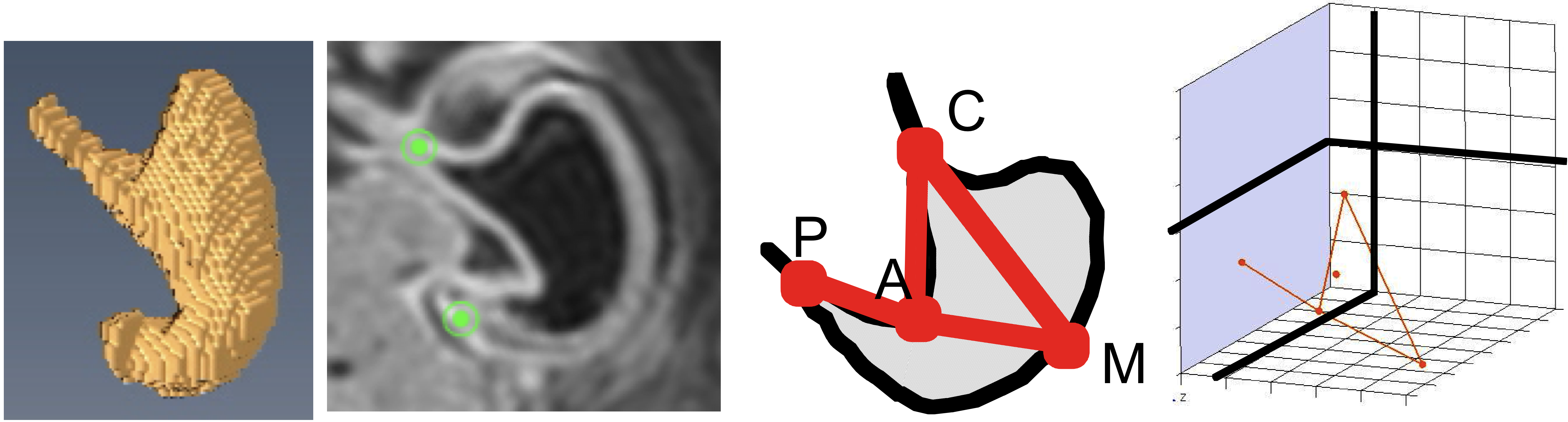
377例の胚子MR画像を用いて、CS16-23の胃の形態形成と動きを検討
stageごとに特徴的な形態
CS18; 胃角、胃底部の隆起
CS18-20; 胃角は90度程度であったが、それ以降鋭角
CS20; 噴門、幽門の分化がみられた。
大弯(M)の3次元的な動き(M), は噴門(C)、幽門(P)の動きと大きく異なる。
C、PはCS16-23の間正中矢状面上にほぼ存在
Mは尾側、左側にCS22まで大きく移動
CPは左右軸を中心に回転
胃の最大平面CPMはおもに頭尾軸を中心に回転
胃の偏位とdifferential growthにより胃は左側、尾側に移動するように見えると推察
CS22の胃; 左から、胃の立体像、 最大断面像、解剖学的観察点、空間座標内の表示
本研究の立体画像元データの一部はMorphoMuseuMに受諾されました。
20. Nako A, Kaigai N, Shiraki N, Yamada S, Uwabe C, Kose K, Takakuwa T, 3D models related to the publication: Morphogenesis of the stomach during the human embryonic period, MorphoMuseuM, in press
ABSTRACT
The stomach develops as the local widening of the foregut after Carnegie stage (CS) 13 that moves in a dramatic and dynamic manner during the embryonic period. Using the magnetic resonance images of 377 human embryos, we present the morphology, morphometry, and three-dimensional movement of the stomach during CS16 and CS23. The stomach morphology revealed stage-specific features. The angular incisura and the cardia were formed at CS18. The change in the angular incisura angle was approximately 90° during CS19 and CS20, and was <90° after CS 21. The prominent formations of the fundus and the pylorus differentiate at around CS20. Morphometry of the stomach revealed that the stomach gradually becomes “deflected” during development. The stomach may appear to move to the left laterally and caudally due to its deflection and differential growth. The track of the reference points in the stomach may reflect the visual three-dimensional movement. The movement of point M, representing the movement of the greater curvature, was different from that of points C (cardia) and P (pyloric antrum). The P and C were located just around the midsagittal plane in all the stages observed. Point M moved in the caudal-left lateral direction until CS22. Moreover, the vector CP does not rotate around the dorsoventral axis, as widely believed, but around the transverse axis. The plane CPM rotated mainly around the longitudinal axis. The data obtained will be useful for prenatal diagnosis in the near future.
Articular cartilage is roughly separated into three areas: the tangential, middle, and deep zones. The structure and molecular components of an additional important zone, the most superficial zone (MSZ), which directly faces the joint cavity, have yet to be conclusively elucidated. The purpose of the present study was to use multiple methods to study the MSZ in order to determine its structure.
Materials and methods
Knees from 16 pigs (age, 6 months) were used. Full-thickness cartilage specimens were harvested from the femoral groove. The MSZ was observed using light microscopy, transmission electron microscopy (TEM), and scanning electron microscopy (SEM) in combination with histochemical and immunohistochemical methods.
Results
The combined findings from the three different observational methods indicate that the MSZ is subdivided into three layers. Among these three layers, collagen subtypes I, II, and III are present in the innermost (third) layer of the MSZ. Beneath the third layer, type II collagen is the predominant type, with small amounts of type III collagen. This layer beneath the third layer is considered to be the tangential layer.
Conclusions
Our observations indicate that the MSZ is subdivided into three layers. Further analysis of the molecular components in each layer may improve our understanding of the structure of the articular surface.
2012年3月に出版しましたthe Human Embryo内のreview, ”Developmental Anatomy of the Human Embryo – 3D-Imaging and Analytical Techniques” ー3次元イメージ解析技術を応用したヒト胚子発生解剖ーのダウンロード回数が1年間で1500を超えました。アメリカ、インド、中国、イタリア等多くの国々からアクセスが有り、月100以上のダウンロードがありました。私たちの研究活動が世界に発信できることは喜ばしいことです。
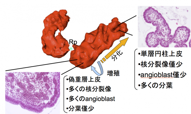
The morphological and histological changes of the choroid plexus (CP) during Carnegie stage (CS) 18 and CS23 were presented, based on magnetic resonance imaging data and histological serial section of human embryos from the Kyoto Collection of Human Embryos. The primordium of the CP was initially detected as a small lump at CS19 that grew caudally, so that the CP became crescent shaped. It developed in all directions after CS21, as the dorsal and frontal growth also became prominent. The CP formed a number of undulating surfaces at CS20, irregular bulges at CS21, and then three large clusters with two deep fissures on the caudal surface at CS23. The mean volume of the CP was 0.282±0.141 mm3 at CS19; it reached 16.8±8.77 mm3 at CS23. Additionally, the histology was different depending on the regions of the CP at all stages after CS20. The epithelium and angioblasts in the center of the stroma were proliferated in the proximal region, whereas the epithelium was differentiated and lobulated in the distal region where the blood vascular system was organized. The histological differentiation was mapped on the CP reconstructed from histological serial sections. The data suggested the correlation between morphological information obtained from magnetic resonance data sets and distribution of the differentiation. With the help of morphological analysis and histological findings, we have been able to categorize each CP into specific stages. These findings will be useful in clinical evaluation of development during the embryonic period.
ヒトの肝臓の形態形成についての論文 (Hirose et al) がAnatomical Recordsの Highlights記事で紹介されました.
AR Highlights
Embryonic Liver Morphology and Morphometry by Magnetic Resonance Microscopic Imaging by Ayumi Hirose, Takashi Nakashima, Shigehito Yamada, Chigako Uwabe, Katsumi Kose, and Tetsuya Takakuwa. Anat Rec 295:51–59
The liver plays an important role in the development of organs in the prenatal period. However, morphological and morphometric features of the liver during the early embryonic period are not well understood. Recent advances in medical imaging have enabled earlier assessment of human development in the first trimester. The authors carried out external morphologic and morphometric analysis of the liver during this period using a superparallel magnetic resonance microscope to image embryos obtained from the Kyoto Collection. They determined the external morphology as well as quantitative morphometry of the embryonic liver. They also found that development of the liver was greatly affected by adjacent organs and tissues. The data from this study provide a better understanding of liver development as well as morphogenesis of nearby organs. The authors’ results could also be used as a reference for clinical evaluation in the early stage of gestation, and this could be useful in fetal medicine and prenatal diagnosis. The authors predict that further improvement in imaging modalities will enable more precise detection of the intrahepatic vascular system.


")

")
")