アクセスカウンター

2024年11月
 123
45678910
11121314151617
18192021222324
252627282930  

肝臓形成不全でも生理的臍帯ヘルニアは起きる: Anat Recに掲載

胚子期、消化管は大きい肝臓に押し出され臍帯内に脱出すると言われています。この機序は、発生の大家Mall博士が19世紀末に提唱し、今多くの教科書に記されています。そうしたら、肝臓形成不全で肝臓が小さい胚子、肝臓がない胚子ではどうなのでしょうか? 

  • 本研究では、消化管臍帯ヘルニアが肝臓の容積に関係なく発生することを示す標本を複数提示することで、発生の大家の提示した説に異議を唱えました。

Kanahashi T, Yamada S, Yoneyama A, Takakuwa T. Relationship Between Physiological Umbilical Herniation and Liver Morphogenesis During the Human Embryonic Period: A Morphological and Morphometric Study. Anat Rec 2019, 302, 1968-1976. doi: 10.1002/ar.24149.

肝臓無形成でも生理的臍帯ヘルニアがみられる

ABSTRACT

It is widely hypothesized that physiological umbilical herniation (PUH) in humans occurs, because the liver occupies a large space in the abdominal cavity, which pushes the intestine into the extraembryonic coelom during the embryonic period. We have recently shown the presence of the intestinal loop in the extraembryonic coelom in embryos with liver malformation. Here, we analyzed the relationship between the liver and the PUH at Carnegie stage 21 of four embryos with liver malformation, including two with hypogenesis (HY1, HY2) and two with agenesis (AG1, AG2), using phase-contrast X-ray computed tomography and compared them with two control embryos. The intestinal loop morphology in the malformed embryos differed from that in the control embryos, except in HY1. The length of the digestive tract in the extraembryonic coelom of the embryos with liver malformation was similar to or longer than that of the controls. The rate of intestinal loop lengthening in the extraembryonic coelom compared with that of the total digestive tract in all embryos with liver malformation was similar to or higher than that of the controls. The estimated total abdominal cavity volume in the embryos with liver malformation was considerably smaller than that of the controls, while the intestinal volume was similar. The cardia and proximal portion of the pancreas connecting to the duodenum were located at almost identical positions in all the embryos, whereas other parts of the upper digestive tract deviated in the embryos with abnormal livers. Thus, our results provided evidence that PUH occurred independently of liver volume.

鈴木さんの修士論文がPLoS ONEに掲載

大腿骨の形態形成

鈴木さんの修士論文がPLoS ONEに受諾されました。

軟骨形成から軟骨内骨化の進む時期の大腿骨の形態形成について外観、内部の変化を位相CT, MRIを用いて解析しました。

  • 軟骨性大腿骨は、CS 18 で初めて観察される
  • 主要な解剖学的ランドマークは、骨化が開始する前(CRL<40mm)に形成
  • 信号強度が高い骨化の開始部位は、しだいに骨幹端軟骨板になる可能性がある骨幹端に限定される
  • 骨化部位の長さ /大腿骨の全長はCRL 40 -75 mmで急速に増加し、CRL が 75 mm で中程度に増加。
  • 軟骨管は、遠位骨端(CRL、75 mm)よりも近位骨端(CRL、62 mm)で早期に発生
  • 骨化後の大腿骨形状の変化は、初期骨化時とその前後に限定的
  • ただし、大腿骨頸部の前傾および大腿骨頭のねじれは、胎児期に連続的に変化

<修士論文の概要>

Suzuki Y, Matsubayashi J, Ji X, Yamada S, Yoneyama A, Imai H, Matsuda T, Aoyama T, Takakuwa T Morphogenesis of the femur at different stages of normal human development, PLoS ONE, 14(8): e0221569. https://doi.org/10.1371/journal. pone.0221569

Abstract

The present study aimed to better characterize the morphogenesis of the femur from the embryonic to the early fetal periods. Sixty-two human fetal specimens (crown–rump length [CRL] range: 11.4–185 mm) from the Kyoto Collection were used for this study. The morphogenesis and internal differentiation process of the femur were analyzed in 3D using phase-contrast X-ray computed tomography and magnetic resonance imaging. The cartilaginous femur was first observed at Carnegie stage 18. Major anatomical landmarks were formed prior to the initiation of ossification at the center of the diaphysis (CRL, 40 mm), as described by Bardeen. The region with very high signal intensity (phase 5 according to Streeter’s classification; i.e., area described as cartilage disintegration) emerged at the center of the diaphysis, which split the region with slightly low signal intensity (phase 4; i.e., cartilage cells of maximum size) in fetuses with a CRL of 40.0 mm. The phase 4 and phase 5 regions became confined to the metaphysis, which might become the epiphyseal cartilage plate. Femur length and ossified shaft length (OSL) showed a strong positive correlation with CRL. The OSL-to-femur length ratio rapidly increased in fetuses with CRL between 40 and 75 mm, which became moderately increased in fetuses with a CRL of ≥75 mm. Cartilage canal invasion occurred earlier at the proximal epiphysis (CRL, 62 mm) than at the distal epiphysis (CRL, 75 mm). Morphometry and Procrustes analysis indicated that changes in the femur shape after ossification were limited, which were mainly detected at the time of initial ossification and shortly after that. In contrast, femoral neck anteversion and torsion of the femoral head continuously changed during the fetal period. Our data could aid in understanding the morphogenesis of the femur and in differentiating normal and abnormal development during the early fetal period.

TMIMS symposiumで発表

大脳subplate層の形成

20th TMIMS International Symposium “Principles of Neocortical Development and Evolution” (2019.7.30, Tokyo)で発表しました

Terashima M, Ishikawa A, Yamada S, Takakuwa T, Morphogenesis of the layer structure of cerebral cortex during human late-embryonic period

第59回先天異常学会で発表

第59回先天異常学会で、発表しました(2019.7.26-28, 名古屋)The 13th World Congress of the International Cleft Lip and Palate Foundation -CLEFT 2019-と合同開催でした。

Nohara A, Owaki N, Manesco C, Katsube M, Yamada S, Imai H, Matsuda T, Yoneyama A, Takakuwa T, Relationship between fusion of lateral palatal shelves and growth of Mandible (Meckel’s cartilage)

PUH occurred independently of liver

Takakuwa T. Intestinal loop formation: herniation into the extraembryonic coelom and return to the abdominal coelom (招待講演)

Drastic changes occur during the development of the intestinal loop (IL), including physiological umbilical herniation (PUH) and its return. The present study was designed to analyze such developments three-dimensionally during human embryonic and early fetal period.

Materials and Methods: The software AMIRA was used to analyze the 3D digitalized data (high-resolution MRI, phase-contrast X-ray CT) obtained from the Kyoto Collection.

Results and Discussion: Based on the results of our analysis, the following time line and main features of IL formation were revealed:

Herniate phase (Carnegie stage (CS)14-CS23, Crown-rump length (CRL) < 35 mm): IL rotation was initially observed as a slight deviation of the duodenum and colorectum from the median plane up to CS16. The PUH was noticeable after CS16. The IL displayed a hairpin-like structure, with the superior mesenteric artery (SMA) running parallel to the straight part and the cecum located to the left at CS18. The IL rotated around the SMA only during the early stages (until CS19). The IL gradually moved away, running transversely after CS19. Embryos with liver malformation showed PUH, which indicated that PUH occurred independent of liver volume.

Transition phase (CRL = 37, 41, and 43 mm): Intestinal return began from proximal to distal part in samples with CRL of 37 mm. The cecum returned before the distal end of the small intestine (ileum) in samples with CRLs of 41 and 43 mm.

Return phase: The cecum immediately reached its final position in the right lower quadrant of the abdomen (the adult position). The anti-clockwise “en-bloc rotation” described by descent and fixation of the cecum in the abdominal cavity may not exist. A rapid increase in the space available for the intestine in the abdominal coelom that exceeded the intestinal volume in the extraembryonic coelom was observed. The height of the umbilical ring increased in a stepwise manner between the transition and return phases and its height in the return phase was comparable to or higher than that of the hernia tip during the herniation phase. We speculated that the space is generated to accommodate the herniated portion of the intestine, similar to the intestine wrapping into the abdominal coelom as the height of the umbilical ring increases.

Conclusion: The data obtained in the present study demonstrate the precise timeline of IL formations, which indicate several points of discrepancy in the results of previous studies.

長田さん、八田くんの卒研がCongenital Anomaliesに受諾(2)

長田さん、八田くんの卒研の一部がCongenital Anomaliesに受諾されました。

胎児期に消化管が臍帯から腹腔に還納されるときに、回盲部がどのように移動するかを3D座標をとり検討しました。

腹腔に戻った回盲部は右下腹部にまっすぐ進んでいく

右上腹部に還納し下腹部に移動するという説も、以前は信じられていましたが、正しくないようです。

42. Nagata A, Hatta S, Imai H, Yamada S, Takakuwa T. Position of the cecum in the extraembryonic and abdominal coelom in the early fetal period. Congenit Anom 2020, 60 (3) 87-88. doi: 10.1111/cga.12348.

第124回 日本解剖学会で発表

第124回日本解剖学会で発表いたしました。(新潟, 3/27-3/29)

ポスター

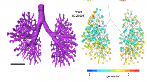
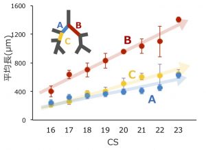
ヒト胚子期における気管支分岐形成の3次元的解析; 藤井瀬菜、村中太河、松林潤、米山明男、武田徹、兵藤一行、山田重人、高桑徹也

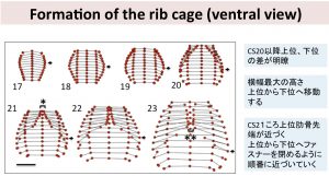
胎児期初期における骨盤形成の解析; 金橋 徹、奥村 美咲、今井 宏彦、山田 重人、山本   憲、富樫 かおり、高桑 徹也

口演

ヒト胎児の顔面骨格形成とそれに必要な因子について; 勝部 元紀、山田 重人、山口 豊、高桑 徹也、山本 憲、斉藤 篤、清水 昭伸、今井 宏彦、鈴木 茂彦

シンポジウム

幾何学的形態測定学法を用いたヒト胎児脳の成長過程の解析; 山口豊、勝部元紀、上部千賀子、巻島美幸、山本憲、今井宏彦、高桑徹也、富樫かおり、山田重人

第18回日本再生医療学会総会で発表しました

第18回日本再生医療学会総会で発表しました (2019.3.21@神戸市)

再生医療において、重要な課題である細胞や組織の生死評価を行いました。3次元構造をとる組織は基質を有し、遊離した細胞よりも測定方法が困難であるとされていますが、本研究では3Dバイオプリンターを用いて3次元構造の組織体を作製し、組織構成体の摘出手法および生死評価手法について検討しました。(今後は、このような3次元的組織構成体の生死判定は移植前の組織の安全性を測るのみでなく移植後のリハビリテーションの有効性の検討を行う際の組織状態のベースラインの確認にも有用であると考えられます。)

石川葵、田中麻衣、杉山寛恵、池口良輔、高井治美、鳥井蓉子、國富芳博、秋枝静香、青山朋樹、高桑徹也 「神経様3次元組織体の生死判定技術の検討」

長田さん、八田くんの卒業論文がJ Anat に掲載

長田さん、八田くんの卒業論文がJournal of Anatomy に掲載されました。

ヒトの消化管は一時的に臍帯内に脱出しCRL40 mmころ、突然還納します(生理的ヘルニア)。生理的ヘルニアの脱出については論文がある程度ありますが、還納過程については研究がほとんどなく、不明な点が多くありました。今回の解析では、還納途中の3体を含む25体について形態計測的な検討をし以下の知見をえました。

  • 腹腔内での占拠割合は徐々に消化管が上昇、肝臓が減少し急激な変化はみられない
  • 消化管還納終了時、腹腔内で使用可能な消化管体積は200mm3で、臍帯内の最大容積25.8mmに比べ十分に大きい
  • ヘルニア期の腸管の最大の高さよりも、還納後の腹壁(臍帯輪)の高さは高い
  • 消化管還納時には、腹腔内へ張力が働く(loop model)という古典的な説に対して、
  • 腹壁がもちあがり消化管を包むことが起きている可能性を提唱(wrapping model)

35. Nagata A, Hatta S, Ji X, Ishikawa A, Sakamoto R, Yamada S, Imai H, Matsuda T, Takakuwa T. Return of the intestinal loop to the abdominal coelom after physiological umbilical herniation in the early fetal period. J Anat, 2019, 234, 456-464.doi: 10.1111/joa.12940.

Abstract

The intestine elongates during the early fetal period, herniates into the extraembryonic coelom, and subsequently returns to the abdominal coelom. The manner of herniation is well-known; however, the process by which the intestinal loop returns to the abdomen is not clear. Thus, the present study was designed to document and measure intestinal movements in the early fetal period in three dimensions to elucidate the intestinal loop return process. Magnetic resonance images from human fetuses whose intestinal loops herniated (herniated phase; n = 5) while returning to the abdominal coelom [transition phase; n = 3, crown–rump length (CRL)] 37, 41, and 43 mm] and those whose intestinal loops returned to the abdominal coelom normally (return phase; n = 12) were selected from the Kyoto Collection. Intestinal return began from proximal to distal in samples with CRL of 37 mm. Only the ileum ends were observed in the extraembryonic coelom in samples with CRLs of 41 and 43 mm, whereas the ceca were already located in the abdominal coeloms. The entire intestinal tract had returned to the abdominal coelom in samples with CRL > 43 mm. The intestinal length increased almost linearly with fetal growth irrespective of the phase (R2 = 0.90). The ratio of the intestinal length in the extraembryonic coelom to the entire intestinal length was maximal in samples with CRLs of 32 mm (77%). This ratio rapidly decreased in three of the samples that were in the transition phase. The abdominal volumes increased exponentially (to the third power) during development. The intestinal volumes accounted for 33–41% of the abdominal volumes among samples in the herniated phase. The proportion of the intestine in the abdominal cavity increased, whereas that in the liver decreased, both without any break or plateau. The amount of space available for the intestine by the end of the transition phase was approximately 200 mm3. The amount of space available for the intestine in the abdominal coelom appeared to be sufficient at the beginning of the return phase in samples with CRLs of approximately 43 mm compared with the maximum intestinal volume available for the extraembryonic coelom in the herniated phase, which was 25.8 mm3 in samples with CRLs of 32 mm. A rapid increase in the space available for the intestine in the abdominal coelom that exceeded the intestinal volume in the extraembryonic coelom generated an inward force, leading to a ‘sucked back’ mechanism acting as the driving force. The height of the hernia tip increased to 8.9 mm at a maximum fetal CRL of 37 mm. The height of the umbilical ring increased in a stepwise manner between the transition and return phases and its height in the return phase was comparable to or higher than that of the hernia tip during the herniation phase. We surmised that the space was generated in the aforementioned manner to accommodate the herniated portion of the intestine, much like the intestine wrapping into the abdominal coelom as the height of the umbilical ring increased.