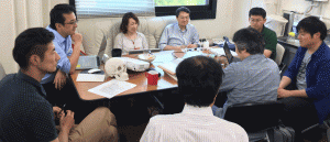
S基盤研究(S)ヒト脳の形態形成から行動生成に至る発達のダイナミクスのメンバーが東京から来られ、研究についてのDiscussionを行いました。
|
||||
|
S基盤研究(S)ヒト脳の形態形成から行動生成に至る発達のダイナミクスのメンバーが東京から来られ、研究についてのDiscussionを行いました。
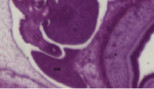
2年ぶりなので演題をたくさん持って行きました。 「先天異常」というのは病理学の大事な分野の一つなのですが、病理学会内で、私たちと類似のことを行っている研究室はないようです。 (口演1) 膝関節の形態形成; EFICを用いた3次元的解析 (示説6) 3次元プリンタを用いたヒト胚子由来立体模型の作製 関節軟骨の最表層は3層の構造で構成される ヒト胚子期における側脳室脈絡叢の形態、組織学的研究 胎児側脳室の形態と長さ計測の有用性 ヒト胚子期の胃の形態形成と3次元的な動き ヒト胎児の肝臓形態形成 第5回病理学技術者講習会(西日本) が平成27年6月27日(土)に行われます。 第9回京都府細胞診ワークショップ、実技講習会
平成27年度 新学術領域研究「多元計算解剖学」シンポジウム (2015.4.30:東京大学)で発表しました。 ヒト器官形成期において分岐構造を有する器官の3次元分枝パターンを解析する
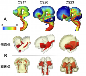
 中島君、片山さん、白石くんの3人の学生や、多くのOffice Assistantの協力を得て、6年かけて解析した論文「ヒト胚子期における脳形態形成」がNeuroimageに受諾されました。また、SupplyのビデオはData in Briefに、3D元データの一部はMorphoMに掲載されました。
13. Shiraishi N, Katayama A, Nakashima T, Yamada S, Uwabe C, Kose K, Takakuwa T, Morphology and morphometry of the human embryonic brain: A three-dimensional analysis, NeuroImage, 2015, 115, 96-103, 10.1016/j.neuroimage.2015.04.044, (概要), [OpenAccess] 12. Shiraishi N, Katayama A, Nakashima T, Yamada S, Uwabe C, Kose K, Takakuwa T, Three-dimensional morphology of the human embryonic brain, Data in Brief, 2015, 4, 116-118, 10.1016/j.dib.2015.05.001 [OpenAccess] 15. Shiraishi N, Katayama A, Nakashima T, Shiraki N, Yamada S, Uwabe C, Kose K, Takakuwa T, 3D model related to the publication: Morphology of the human embryonic brain and ventricles, MorphoMuseuM 1 (3)-e3. doi: 10.18563/m3.1.3.e3. [OpenAccess] AbstractThe three-dimensional dynamics and morphology of the human embryonic brain have not been previously analyzed using modern imaging techniques. The morphogenesis of the cerebral vesicles and ventricles was analyzed using images derived from human embryo specimens from the Kyoto Collection, which were acquired with a magnetic resonance microscope equipped with a 2.35-T superconducting magnet. A total of 101 embryos between Carnegie stages (CS) 13 and 23, without apparent morphological damage or torsion in the brain ventricles and axes, were studied. To estimate the uneven development of the cerebral vesicles, the volumes of the whole embryo and brain, prosencephalon, mesencephalon, and rhombencephalon with their respective ventricles were measured using image analyzing Amira™ software. The brain volume, excluding the ventricles (brain tissue), was 1.15 ± 0.43 mm3 (mean ± SD) at CS13 and increased exponentially to 189.10 ± 36.91 mm3 at CS23, a 164.4-fold increase, which is consistent with the observed morphological changes. The mean volume of the prosencephalon was 0.26 ± 0.15 mm3 at CS13. The volume increased exponentially until CS23, when it reached 110.99 ± 27.58 mm3. The mean volumes of the mesencephalon and rhombencephalon were 0.20 ± 0.07 mm3 and 0.69 ± 0.23 mm3 at CS13, respectively; the volumes reached 21.86 ± 3.30 mm3 and 56.45 ± 7.64 mm3 at CS23, respectively. The ratio of the cerebellum to the rhombencephalon was approximately 7.2% at CS20, and increased to 12.8% at CS23. The ratio of the volume of the cerebral vesicles to that of the whole embryo remained nearly constant between CS15 and CS23 (11.6–15.5%). The non-uniform thickness of the brain tissue during development, which may indicate the differentiation of the brain, was visualized with surface color mapping by thickness. At CS23, the basal regions of the prosencephalon and rhombencephalon were thicker than the corresponding dorsal regions. The brain was further studied by the serial digital subtraction of layers of tissue from both the external and internal surfaces to visualize the core region (COR) of the thickening brain tissue. The COR, associated with the development of nuclei, became apparent after CS16; this was particularly visible in the prosencephalon. The anatomical positions of the COR were mostly consistent with the formation of the basal ganglia, thalamus, and pyramidal tract. This was confirmed through comparisons with serial histological sections of the human embryonic brain. The approach used in this study may be suitable as a convenient alternative method for estimating the development and differentiation of the neural ganglia and tracts. These findings contribute to a better understanding of brain and cerebral ventricle development. HighlightsThree-dimensional morphogenesis of the human embryonic brain was presented from MRI.• The volume of three brain vesicles and ventricles were measured at 101 embryos.• The brain volume exponentially increased 164.4-fold from Carnegie stage 13 to 23.• The volume ratio of the brain to whole embryo remained nearly constant.• The non-uniform thickness of the brain tissue during development was 3D visualized. 遠藤さんの卒業論文「ヒト胚子脾臓の形態形成」がAnat Recに掲載されました。 脾臓は、左上腹部の胃の後方に位置する腹腔内の代表的な器官です。脾臓は成人では。成人の脾臓の主な役割は、血液の濾過・不要な物質の除去と免疫系としてのリンパ球の産生です。ヒトにおける初期発生過程はこれまでほとんど知られていませんでした。本研究では、脾臓の形態形成、および脾臓内外の血管の形成過程についてCarnegie stageごとに記載しました。 
11.Endo A, Ueno S, Yamada S, Uwabe C, Takakuwa T, Morphogenesis of the spleen during the human embryonic period, Anatomical Rec, 2015, 298, 820-826, doi: 10.1002/ar.23099 ABSTRACTWe aimed to observe morphological changes in the spleen from the emergence of the primordium to the end of the embryonic period using histological serial sections of 228 samples. Between Carnegie stages (CSs) 14 and 17, the spleen was usually recognized as a bulge in the dorsal mesogastrium (DM), and after CS 20, the spleen became apparent. Intrasplenic folds were observed later. A high-density area was first recognized in 6 of the 58 cases at CS 16 and in all cases examined after CS 18. The spleen was recognized neither as a bulge nor as a high-density area at CS 13. The mesothelium was pseudostratified until CS 16 and was replaced with high columnar cells and then with low columnar cells. The basement membrane was obvious after CS 17. The mesenchymal cells differentiated from cells in the DM, and sinus formation started at CS 20. Hematopoietic cells were detected after CS 18. The vessels were observed at CS 14 in the DM. Hilus formation was observed after CS 20. The parallel entries of the arteries and veins were observed at CS 23. The rate of increase in spleen length in relation to that of stomach length along the cranial-caudal direction was 0.51 ± 0.11, which remained constant during CSs 19 and 23, indicating that their growths were similar. These data may help to better understand the development of normal human embryos and to detect abnormal embryos in the early stages of development. 竹谷さんの卒業論文が、 Congenital Anomalies 55巻(2015)に掲載されました。 本論文の内容は第104回日本病理学会総会で発表しました。
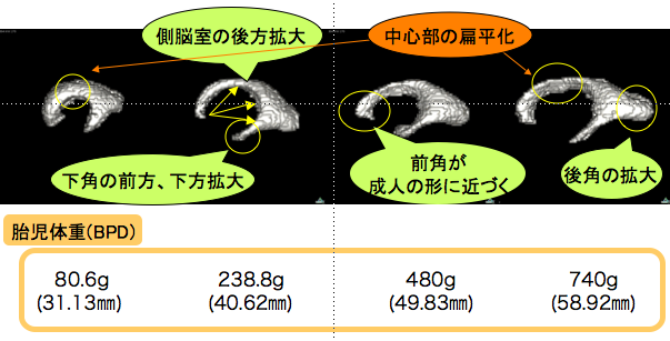
 Taketani K, Yamada S, Uwabe C, Okada T, Togash K, Takakuwa T, Morphological features and length measurements of fetal lateral ventricles at 16–25 weeks of gestation by magnetic resonance imaging, Congenit Anom (Kyoto), 2015, 55, 99-102. doi: 10.1111/cga.12076 AbstractNormal growth of the lateral ventricles (LVs) was characterized three-dimensionally using magnetic resonance imaging (MRI) data from 16 human fetuses at 16–25 weeks of gestation. The LV was differentiated into four primary regions, the anterior horn, central parts, posterior horn, and inferior horn, at 16 weeks of gestation. The LV changed shape mainly by elongation and narrowing, which corresponded to the external and internal growth of the surrounding cerebrum. Six length parameters measured in the LV correlated with biparietal diameter by simple regression analysis (R2 range, 0.56–0.93), which may be valuable for establishing a standardized prenatal protocol to assess fetal well-being and development across intrauterine periods. No correlation was found between biparietal diameter and LV volume (R2 = 0.13). |
||||