 第25回日本基礎理学療法学会学術大会で発表しました。2020.12.12-13 @仙台市(on line開催)
石川葵、田中麻衣、杉山寛恵、池口良輔、高井治美、鳥井蓉子、國富芳博、秋枝静香、谷間桃子、青山朋樹: 神経様3次元組織体の生死判定技術の検討
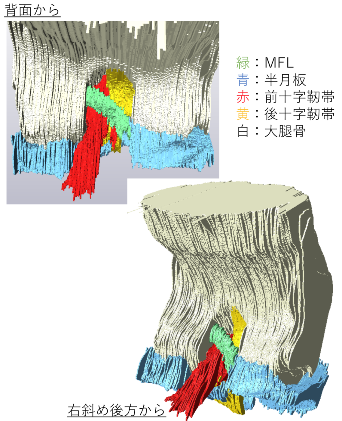
石田かのん、谷間桃子、石川葵、高石亮太、高桑徹也、青山朋樹: ラット膝関節の発生と 半月大腿靭帯の形態形成
|
||||
  |
石川葵、田中麻衣、杉山寛恵、池口良輔、高井治美、鳥井蓉子、國富芳博、秋枝静香、谷間桃子、青山朋樹: 神経様3次元組織体の生死判定技術の検討 石田かのん、谷間桃子、石川葵、高石亮太、高桑徹也、青山朋樹: ラット膝関節の発生と 半月大腿靭帯の形態形成 季さんの論文がAnat Recに掲載されました。  胎児期初期の臍帯ヘルニアの還納と腹壁(腹直筋)の形成との関連性について検討しました。
Ji X, Ishikawa A, Nagata A, Yamada S, Imai H, Matsuda T, Takakuwa T, Relationship between rectal abdominis muscle position and physiological umbilical herniation and return: a morphological and morphometric study. Anat Rec 2020, 303, 12, 3044-3051. doi: 10.1002/ar.24486 受諾は1年前だったのですが、ようやくpublishされました。 AbstractThe herniation of the intestinal loop (IL) in the extraembryonic coelom and its return to abdominal cavity is in parallel with the formation of the rectal abdominis muscle (RAM). Using high-resolution magnetic resonance imaging data of human fetuses (n = 19, CRL22-69 mm; stored at Kyoto Collection), this study aimed to analyze the relationship between the development of RAM and phase of IL herniation. The RAM runs at the lateral part of the abdominal wall in the small samples in the herniation phase. The position was shifted to the midline area in the larger samples in the herniation phase. According to fetal growth, the caudal ends of the muscles extended along the umbilical ring towards the pubis, though the caudal part of the RAMs were thin and faint in most of the samples. Length measurements related with the growth of the abdominal wall including RAM and abdominal circumference showed positive correlation with fetal growth. On the contrary, diastasis of RAMs and the width and area of the umbilical ring were almost constant according to fetal growth. Such morphometric value showed no obvious changes regardless of the phases of herniation. The ratio of the width and diastasis of the RAMs to the circumference was decreased, indicating that the closure of the ventral body wall was influenced by growth differences. The present data indicate that the formation of the abdominal wall including RAM is independent of the phase of IL herniation, whether in the extraembryonic coelom or in the abdominal cavity.  西谷さんの修士論文、鳥居さんの卒業論文;胎児心臓のDT-MRIによる解析がJAHAに掲載されました。
本研究成果は、京都大学HP(トピックス)でも紹介されました。 ヒト胎児心臓の心筋線維方向を追跡 -受精後8週の心筋線維は成人と同じ配列をする- 外部リンク[京都大学HP(トピックス)] 2020/10/13 医療NEWSにて,ヒト胎児の心臓、受精後8週で心筋線維の配列が成人と同様であることが判明-京大ほか が紹介されました. 46. Nishitani S, Torii N, Imai H, Haraguchi R, Yamada S, Takakuwa T, Development of helical myofiber tracts in the human fetal heart: Analysis of myocardial fiber formation in the left ventricle from the late human embryonic period using diffusion tensor magnetic resonance imaging. Journal of the American Heart Association, 2020, 19(9) doi:10.1161/JAHA.120.016422 AbstractBackgroundDetection of the fiber orientation pattern of the myocardium using diffusion tensor magnetic resonance imaging lags ≈12 weeks of gestational age (WGA) behind fetal myocardial remodeling with invasion by the developing coronary vasculature (8 WGA). We aimed to use diffusion tensor magnetic resonance imaging tractography to characterize the evolution of fiber architecture in the developing human heart from the later embryonic period. Methods and ResultsTwenty human specimens (8–24 WGA) from the Kyoto Collection of Human Embryos and Fetuses, including specimens from the embryonic period (Carnegie stages 20–23), were used. Diffusion tensor magnetic resonance imaging data were acquired with a 7T magnetic resonance system. Fractional anisotropy and helix angle were calculated using standard definitions. In all samples, the fibers ran helically in an organized pattern in both the left and right ventricles. A smooth transmural change in helix angle values (from positive to negative) was detected in all 16 directions of the ventricles. This feature was observed in almost all small (Carnegie stage 23) and large samples. A higher fractional anisotropy value was detected at the outer side of the anterior wall and septum at Carnegie stage 20 to 22, which spread around the ventricular wall at Carnegie stage 23 and in the early fetal samples (11–12 WGA). The fractional anisotropy value of the left ventricular walls decreased in samples with ≥13 WGA, which remained low (≈0.09) in larger samples. ConclusionsFrom the human late embryonic period (from 8 WGA), the helix angle arrangement of the myocardium is comparable to that of the adult, indicating that the myocardial structure blueprint, organization, and integrity are already formed. 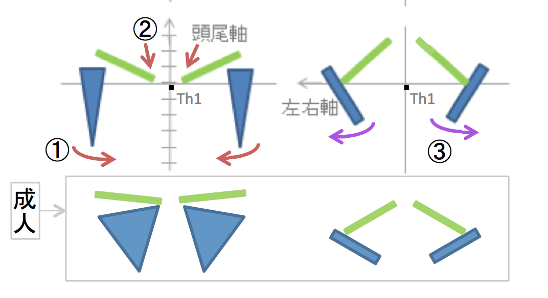 田中さん、坂本さんの卒業研究がPLoS ONEに掲載されました。胎児期の上肢帯の形態形成、位置の変化について詳述しました。
46. Tanaka S, Sakamoto R, Kanahashi T, Yamada S, Imai H, Yoneyama A, Takakuwa T. Shoulder girdle formation and positioning during embryonic and early fetal human development. PLoS ONE 2020, 15(9): e0238225. https://doi.org/10.1371/journal.pone.0238225 AbstractPositional information on the shoulder girdle (the clavicle and scapula) is important for a better understanding of the function of the upper limb in the locomotive system as well as its associated disease pathogenesis. However, such data are limited except for information on the axial position of the scapula. Here, we describe a three-dimensional reconstruction of the shoulder girdle including the clavicle and scapula, and its relationship to different landmarks in the body. Thirty-six human fetal specimens (crown-rump length range: 7.6–225 mm) from the Kyoto Collection were used for this study. The morphogenesis and three-dimensional position of the shoulder girdle were analyzed with phase-contrast X-ray computed tomography and magnetic resonance imaging. We first detected the scapula body along with the coracoid and humeral head at Carnegie stage 18; however, the connection between the body and coracoid was not confirmed at this stage. During development, all landmarks on the shoulder girdle remained at the same axial position except for the inferior angle, which implies that the scapula enlarged in the caudal direction and reached the adult axial position in the fetal period. The scapula body was rotated internally and in the upward direction at the initiation of morphogenesis, but in the fetal period the scapula body was different than that in the adult position. The shoulder girdle was located at the ventral side of the vertebrae at the time of initial morphogenesis, but changed its position to the lateral side of the vertebrae in the late embryonic and fetal periods. Such a unique position of the shoulder girdle may contribute to the stage-specific posture of the upper limb. Adequate internal and upward rotation of the scapula could help in reducing the shoulder width, thereby facilitating childbirth. The data presented in this study can be used as normal morphometric references for shoulder girdle evaluations in the embryonic and fetal periods. 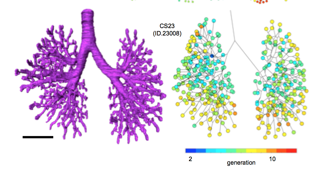 藤井さんの博士論文がJ Anatomy に受諾されました。 詳細な形態変化情報と正確な分岐次数を算出することにより、これまで明らかでなかった胚子期の気管支形成について新たな知見を与えました。
44. Fujii S, Muranaka T, Matsubayash J, Yamada S, Yoneyama A, Takakuwa T. The bronchial tree of the human embryo: an analysis of variations in the bronchial segments. J Anatomy 2020, 237, 311-322. doi: 10.1111/joa.13199. Abstract A classical study has revealed the general growth of the bronchial tree and its variations up to Carnegie stage (CS) 19. In the present study, we extended the morphological analysis CS by CS until the end of the embryonic period (CS23). A total of 48 samples between CS15 and CS23 belonging to the Kyoto Collection were used to acquire imaging data by performing phase-contrast X-ray computed tomography. Three-dimensionally reconstructed bronchial trees revealed the timeline of morphogenesis during the embryonic period. Structures of the trachea and lobar bronchus showed no individual difference during the analyzed stages. The right superior lobar bronchus was formed after the generation of both the right middle lobar bronchus and the left superior lobar bronchus. The speed of formation of the segmental bronchi, sub-segmental bronchi, and further generation seemed to vary among individual samples. The distribution of the end-branch generation among five lobes was significantly different. The median branching generation value in the right middle lobe was significantly low compared with that of the other four lobes, whereas that of the right inferior lobe was significantly larger than that of both the right and left superior lobes. Variations found between CS20 and CS23 were all described in the human adult lung, indicating that variation in the bronchial tree may well arise during the embryonic period and continue throughout life. The data provided may contribute to a better understanding of bronchial tree formation during the human embryonic period.
The bronchial tree of the human embryo: an analysis of (ヒト胚子期の気管支樹:区域気管支の多様性の検討) 気管支の中枢部分は葉気管支と区域気管支から成るが、多くの成人肺の解剖学的研究においてこの典型的な構造から逸脱した多様な分岐構造が報告されている。これまでに気管支の多様性が生じる過程を明らかにした研究はない。本研究は、Carnegie stage (CS) 15〜23のヒト胚子標本の位相CT画像を用いて作成した気管支樹の三次元立体像に解析を加えることで、胚子期における気管支の多様性を検討した。形態観察および区域気管支までの同定をおこない、各葉の分岐次数を算出した。CS15からCS16の観察結果より、右上葉は右中葉と左上葉に遅れて形成することが示された。CS17~19において、区域気管支と亜区域気管支が、同一ステージの個体間で形成の程度にばらつきを示しながら形成していた。分岐次数は各葉にてCSの進行とともに増加した。右中葉の分岐次数の中央値は他の4葉と比較して有意に低かった。CS20~23の気管支樹における区域気管支について、全5葉にて14種類の形態を同定した。本研究にて同定した形態は、すべて成人の気管支にて報告のある形態に含まれていた。よって、区域気管支の多様な構造は胚子期に決定する可能性が示唆された。本研究は、詳細な形態変化情報と正確な分岐次数を算出することにより、これまで明らかでなかった胚子期の気管支形成について新たな知見を与えたものと考える。
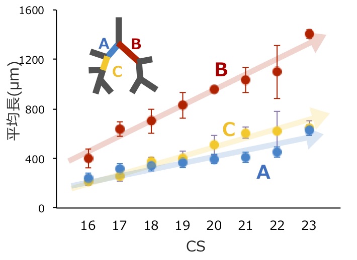
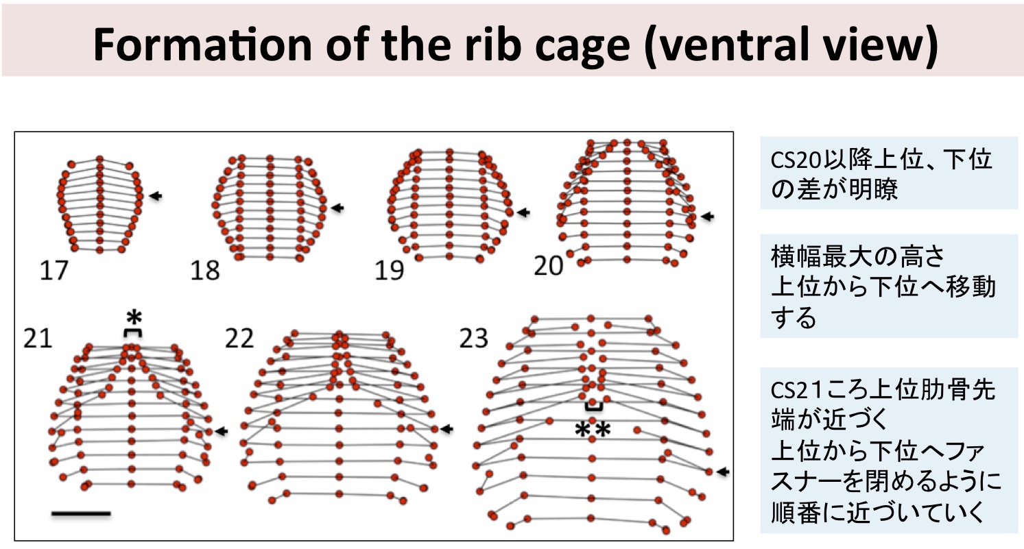
博士号を取得して 寄稿の機会をいただきありがとうございます。2017年に京都大学大学院医学研究科人間健康科学系専攻へ社会人博士として入学し、2020年11月に博士号を取得することができました。ここでは個人的に印象深かった出来事を著し、博士号取得の挨拶に代えさせていただきます。 初めて科学雑誌に論文を投稿したのは2019年11月、査読の返答は12月の終わり頃に届きました。査読者のコメントにより投稿内容のおよそ半分を入れ替えることとなり、査読へ対応するための原稿の修正、後輩の修士論文執筆に向けた解析の支援と、その年の年末年始は博士課程在学中でも特に充実した時間であったと記憶しています。修正原稿の投稿から約ひと月後の早朝、指導教員の簡潔な2行ほどのメールにより、受諾の知らせが届きました。簡潔な内容に驚きつつ、顔を合わせた際にはどんなありがたいお言葉をいただけるのだろうと、淡い期待を胸に、底冷え残る京都の朝、研究室へ向かいました。しかし、教授は学位申請の手続きを早めに行うよう勧めたのち、受諾論文では省いたデータを改めて論文にまとめる話を切り出されました。このとき、職業研究者というものを多少なりとも理解したように思います。 当時省いたデータは、査読にて指摘を受けた部分を補強する解析を行ってまとめ直し、こちらも論文として受諾されました。そして、現在は次の解析へと目を向けています。最後に、このように博士課程で得た成果を論文として投稿することができたのは、指導教員ならびに共同研究者の皆様のご尽力の賜物であります。この場を借りて厚く御礼申し上げます。 Fujii S, Muranaka T, Matsubayash J, Yamada S, Yoneyama A, Takakuwa T. The bronchial tree of the human embryo: an analysis of variations in the bronchial segments. J Anatomy 2020, 237, 311-322. doi: 10.1111/joa.13199. Matsubayashi J, Okuno K, Fuji S, Ishizu K, Yamada S, Yoneyama A, Takakuwa T. Human embryonic ribs all progress through common morphological forms irrespective of their position on the axis, Dev Dyn 2019, 248, 1257-1263, doi: 10.1002/dvdy.107 Okuno K, Ishizu K, Matsubayashi J, Fujii S, Sakamoto R, Ishikawa A, Yamada S, Yoneyama A, Takakuwa T. Rib cage morphogenesis in the human embryo: A detailed three-dimensional analysis. Anat Rec 2019, 302, 2211-2223, doi: 10.1002/ar.24226 Ishiyama H, Ishikawa A, Kitazawa H, Fujii S, Matsubayashi J, Yamada S, Takakuwa T, Branching morphogenesis of the urinary collecting system in the human embryonic metanephros, PLoS ONE 13(9): e0203623. doi: 10.1371/journal.pone.0203623
新型コロナウイルス感染症の影響でon line 学術開催となりました。 金橋 徹、山田重人、米山明男、高桑徹也, ヒト胚子期に起こる生理的臍帯ヘルニアの発生要因‐肝形成との関連に基づいた形態及び形態計測学的検討 藤井 瀬菜, 村中 太河, 松林 潤, 米山 明男, 兵藤 一行, 山田 重人, 高桑 徹也, ヒト胚子期における気管支の非対称性の定量的検討 共同研究者の勝部先生、東島先生が奨励賞を受賞しました。おめでとうございます。 勝部 元紀 先生
高桑徹也、山田重人、米山明男. ヒト胚子肋骨の形態形成は、共通の肋骨の形状変化のどのあたりに相当するかという尺度で示すことができる |
|||