35. Nagata A, Hatta S, Ji X, Ishikawa A, Sakamoto R, Yamada S, Imai H, Matsuda T, Takakuwa T. Return of the intestinal loop to the abdominal coelom after physiological umbilical herniation in the early fetal period. J Anat, 2019, 234, 456-464.doi: 10.1111/joa.12940.
Abstract
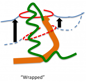
The intestine elongates during the early fetal period, herniates into the extraembryonic coelom, and subsequently returns to the abdominal coelom. The manner of herniation is well-known; however, the process by which the intestinal loop returns to the abdomen is not clear. Thus, the present study was designed to document and measure intestinal movements in the early fetal period in three dimensions to elucidate the intestinal loop return process. Magnetic resonance images from human fetuses whose intestinal loops herniated (herniated phase; n = 5) while returning to the abdominal coelom [transition phase; n = 3, crown–rump length (CRL)] 37, 41, and 43 mm] and those whose intestinal loops returned to the abdominal coelom normally (return phase; n = 12) were selected from the Kyoto Collection. Intestinal return began from proximal to distal in samples with CRL of 37 mm. Only the ileum ends were observed in the extraembryonic coelom in samples with CRLs of 41 and 43 mm, whereas the ceca were already located in the abdominal coeloms. The entire intestinal tract had returned to the abdominal coelom in samples with CRL > 43 mm. The intestinal length increased almost linearly with fetal growth irrespective of the phase (R2 = 0.90). The ratio of the intestinal length in the extraembryonic coelom to the entire intestinal length was maximal in samples with CRLs of 32 mm (77%). This ratio rapidly decreased in three of the samples that were in the transition phase. The abdominal volumes increased exponentially (to the third power) during development. The intestinal volumes accounted for 33–41% of the abdominal volumes among samples in the herniated phase. The proportion of the intestine in the abdominal cavity increased, whereas that in the liver decreased, both without any break or plateau. The amount of space available for the intestine by the end of the transition phase was approximately 200 mm3. The amount of space available for the intestine in the abdominal coelom appeared to be sufficient at the beginning of the return phase in samples with CRLs of approximately 43 mm compared with the maximum intestinal volume available for the extraembryonic coelom in the herniated phase, which was 25.8 mm3 in samples with CRLs of 32 mm. A rapid increase in the space available for the intestine in the abdominal coelom that exceeded the intestinal volume in the extraembryonic coelom generated an inward force, leading to a ‘sucked back’ mechanism acting as the driving force. The height of the hernia tip increased to 8.9 mm at a maximum fetal CRL of 37 mm. The height of the umbilical ring increased in a stepwise manner between the transition and return phases and its height in the return phase was comparable to or higher than that of the hernia tip during the herniation phase. We surmised that the space was generated in the aforementioned manner to accommodate the herniated portion of the intestine, much like the intestine wrapping into the abdominal coelom as the height of the umbilical ring increased.
多元計算解剖学第5回国際シンポジウム(The 5th International Symposium on Multidisciplinary Computational Anatomy)で発表しました。(3/4-5, 九州大学)
A02-KB107 Analysis of Central Nervous System and Skeletal System During Human Early-fetal Period Based on Multidisciplinary Computational Anatomy -Progress Overview FY 2017-FY2018- (PI:Tetsuya Takakuwa) 日本語
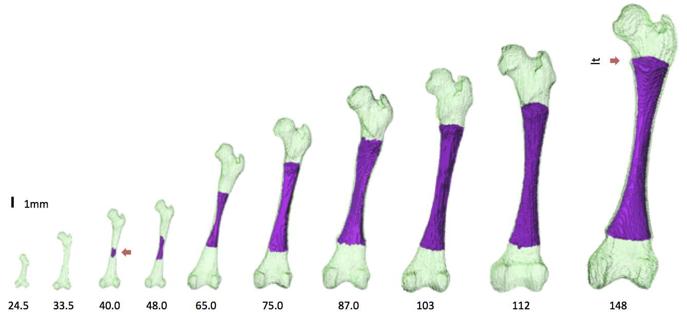
36. Suzuki Y, Matsubayashi J, Ji X, Yamada S, Yoneyama A, Imai H, Matsuda T, Aoyama T, Takakuwa T Morphogenesis of the femur at different stages of normal human development, PLoS One, 14(8): e0221569. https://doi.org/10.1371/journal. pone.0221569
Myocardial fiber histogenesis during human early fetal period using diffusion tensor magnetic resonance imaging(DT-MRI) 高解像度MRI(DTI)を用いたヒト胎児期初期の心筋線維の形成 西谷早織
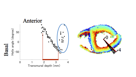
46. Nishitani S, Torii N, Imai H, Haraguchi R, Yamada S, Takakuwa T, Development of helical myofiber tracts in the human fetal heart: Analysis of myocardial fiber formation in the left ventricle from the late human embryonic period using diffusion tensor magnetic resonance imaging. Journal of the American Heart Association, 2020, 19(9), e016422, doi:10.1161/JAHA.120.016422
34. Ishiyama H, Ishikawa A, Kitazawa H, Fujii S, Matsubayashi J, Yamada S, Takakuwa T, Branching morphogenesis of the urinary collecting system in the human embryonic metanephros, PLoS ONE 13(9): e0203623. https://doi.org/10.1371/journal.pone.0203623
Abstract
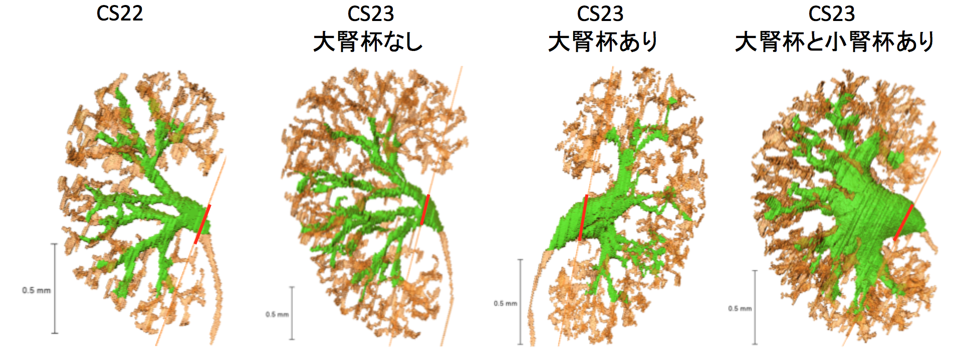
An elaborate system of ducts collects urine from all nephrons, and this structure is known as the urinary collecting system (UCS). This study focused on how the UCS is formed during human embryogenesis. Fifty human embryos between the Carnegie stage (CS) 14 and CS23 were selected from the Kyoto Collection at the Congenital Anomaly Research Center of Kyoto University, Japan. Metanephroses, including the UCS, were segmented on serial digital virtual histological sections. Three-dimensional images were computationally reconstructed for morphological and quantitative analyses. A CS timeline was plotted. It consisted of the 3-D structural morphogenesis of UCS and quantification of the total amount of end-branching, average and maximum numbers of generations, deviation in the metanephros, differentiation of the urothelial epithelium in the renal pelvis, and timing of the rapid expansion of the renal pelvis. The first UCS branching generation occurred by CS16. The average branching generation reached a maximum of 8.74 ± 1.60 and was already the twelfth in CS23. The total end-branching number squared between the start and the end of the embryonic period. UCS would reach the fifteenth branching generation soon after CS23. The number of nephrons per UCS end-branch was low (0.21 ± 0.14 at CS19, 1.34 ± 0.49 at CS23), indicating that the bifid branching occurred rapidly and that the formation of nephrons followed after. The renal pelvis expanded mainly in CS23, which was earlier than that reported in a previous study. The number of nephrons connected to the UCS in the expanded group (246.0 ± 13.2) was significantly larger than that of the pre-expanded group (130.8 ± 80.1) (P < 0.05). The urothelial epithelium differentiated from the zeroth to the third generations at CS23. Differentiation may have continued up until the tenth generation to allow for renal pelvis expansion. The branching speed was not uniform. There were significantly more branching generations in the polar- than in the interpolar regions (P < 0.05). Branching speed reflects the growth orientation required to form the metanephros. Further study will be necessary to understand the renal pelvis expansion mechanism in CS23. Our CS-based timeline enabled us to map UCS formation and predict functional renal capacity after differentiation and growth.
⑦ Miyazaki R, Makishima H, Männer J, Sydow HG, Uwabe C, Takakuwa T, Viebahn C, Yamada S. The Blechschmidt Collection: revisiting specimens from a historical collection of serially sectioned human embryos and fetuses using modern imaging techniques, Congenit Anom, 2018, 58, 152-157, doi: 10.1111/cga.12261
ABSTRACT
Along with the Carnegie Collection in the United States and the Kyoto Collection in Japan, the Blechschmidt Collection (Georg-August-University of Göttingen, Germany) is a major historical human embryo and fetus collection. These collections are of enormous value to human embryology; however, due to the nature of the historical histological specimens, some stains are fading in color, and some glass slides are deteriorating over time. To protect these specimens against such degradation and ensure their future usefulness, we tried to apply modern image scanning and computational reconstruction. Samples of histological specimens of the Blechschmidt Collection were digitized into images using commercial flatbed scanners with a resolution of 4800 pixels per inch. Two specimens were reconstructed into three-dimensional (3D) images by using modern techniques to vertically stack two-dimensional images of the slices into 3D blocks. The larger specimen of crown-rump length (CRL) 64.0 mm, a series of very large histological sections in human embryology, was reconstructed clearly, with its central nervous system segmented before stacking. The smaller specimen of CRL 17.5 mm was also reconstructed into 3D images. The outer surface of the embryo was intact, and its development was classified according to the widely used Carnegie stages (CSs). The CS of the specimen was identified as the later half of CS 20. The invaluable Blechschmidt Collection can be revisited for further research with modern techniques such as digital image scanning and computational 3D reconstruction.
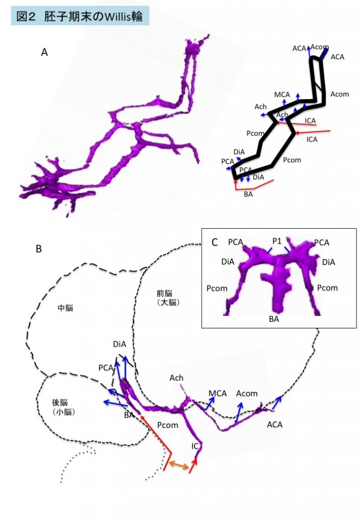
33. Furuichi K, Ishikawa A, Uwabe C, Makishima H, Yamada S, Takakuwa T, Variations of the circle of Willis at the end of the human embryonic period, 2018, 301, 1312-1319, doi:10.1002/ar.23794
ABSTRACT
Variations of the circle of Willis (CW) influence blood supply to the brain and adjacent structures in adults. We examined the formation of the CW in 20 human embryo samples at the end of the embryonic period using 3-D reconstructions of serial histological sections. The CW was closed in all samples, and did not form in a single plane, but was composed of multiple stair-like planes. The artery acutely curved at the caudal part of the CW, namely, at the inlet of the basilar artery and bifurcation of the P1 segment of the posterior cerebral artery (PCA), reflecting flexure of the mesencephalon and diencephalon at this stage. Variations were observed in 17 of 20 samples—only anterior parts (anterior communicating artery [Acom] and anterior cerebral artery [ACA]) in 10 samples, only posterior parts (posterior communicating artery [Pcom]) in one sample, and both anterior and posterior parts in six samples. Variations included the Acom formed as partially duplicated in three samples, duplicated in four, plexiform in three, and no channel as a result of a single azygos ACA in one. The ACA formed as duplicated in two, median ACA in two, and right hypoplasia in one. The Pcom formed in hypoplasia of either side in six samples. Variations observed in this study are similar to those observed in fetuses, neonates, and adults. The P1 segment of PCA was very large in all samples. The present observations indicate that variations in the CW are present from the initiation of CW formation.