アクセスカウンター

2024年11月
 123
45678910
11121314151617
18192021222324
252627282930  

2012年度;修士論文の概要(藤岡瑠音)

Localization of collagen subtypes, which consist the articular surface
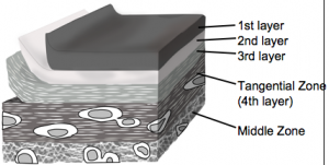
軟骨最表層の構造

軟骨最表層の構造

はじめに:関節軟骨は一般的に細胞の形態、基質の含有により表層、中層、深層の三層に分類される。しかし、関節の潤滑な動きに重要な役割を担うであろう最表層と呼ばれる層が、表層より更に関節面側に存在することは、あまり注目されていない。最表層に関するこれまでの報告では、光学顕微鏡、透過型電子顕微鏡、走査型顕微鏡のいずれかが用いられてきたが、得られた最表層の像は手法により様々で一定化していない。そこで本研究では、光学顕微鏡、透過型電子顕微鏡及び走査型電子顕微鏡を用い、組織学的、免疫組織学的手法を組み合わせた最表層各層の構造・構成成分の比較を行なった。

手法:ブタの膝関節軟骨を用いて、組織化学染色及びⅠ型、Ⅱ型、Ⅲ型コラーゲンに対する免疫染色を行い、光学顕微鏡、及び透過型、走査型電子顕微鏡による観察を行った。

軟骨最表層のSEM像

結果:光学顕微鏡観察により、関節軟骨の最表層はプロテオグリカンに乏しく、方向性をもったコラーゲン性の層を含んでいることが明らかとなった。また、透過型、走査型、及び免疫電子顕微鏡による観察から、最表層は三層構造に分けられた。最表面から第一層、次いで第二層が存在した。第一層は透過型電子顕微鏡では電子密度の高い層として観察でき、無定形物質からなる層であった。第二層は透過型電子顕微鏡でのみ、低電子密度層として確認された。これらの層にはⅠ型、Ⅱ型、Ⅲ型いずれのコラーゲンも含まれていなかった。一方、第三層は透過型電子顕微鏡では識別できないが、線維の密度や走行から走査型顕微鏡で下層と区別して観察された。この第三層はⅠ型、Ⅱ型、Ⅲ型コラーゲンにより構成されていた。第三層の下層は、Ⅱ型コラーゲンから構成されるコラーゲン線維と紡錘形の軟骨細胞からなる、いわゆる表層であった。表層にはⅠ型、Ⅲ型コラーゲンはほとんど分布していなかった。

考察:最表層を構成する三層の中でも、より表面に存在する第一層と第二層では、コラーゲンやプロテオグリカンなどの軟骨構成成分が認められず、第三層はⅠ型、Ⅱ型、Ⅲ型コラーゲンにより構成される事が示された。これらのことから、第一層、二層は第三層以下の軟骨構造とは異なる構成帯であり、第三層は軟骨構成成分であるⅡ型コラーゲンを含むとともに、この層に特異なⅠ型、Ⅲ型のコラーゲンサブタイプを含み、独立した構造体を構成していることが示された。

7. Fujioka R, Aoyama T, Takakuwa T, The layered structure of the articular surface, Osteoarthritis Cartilage, 2013, 21, 1092-1098 doi: 10.1016/j.joca.2013.04.021

脈絡叢の形成; Anat Recに掲載(白石修論)

側脳室内の脈絡叢(正面)

白石くんの修士論文”Morphogenesis of lateral choroid plexus during human embryonic period” -ヒト胚子期における側脳室脈絡叢の形態、組織学的研究-がAnatomical Recordに掲載されました。

  • CS18 – CS23 における脈絡叢 (CP) の形成を検討。
    • CS19 ;CP の原基、尾側に成長した小さな三日月形の塊として検出
    • CS20; 多数の起伏のある表面
    • CS21 で不規則な膨らみを形成、全方向に成長
    • CS23 で尾側表面に 2 つの深裂、 3 つの大きなクラスターを形成
  • 近位領域は未分化な上皮、血管芽細胞が増殖、遠位領域は分化、分葉化した組織を観察
近位領域では未分化な上皮、血管芽細胞が増殖、遠位領域では分化、分葉化した組織がみられる。

6. Shiraishi N, Nakashima T, Yamada S, Uwabe C, Kose K, Takakuwa T, Morphogenesis of lateral choroid plexus during human embryonic period, Anat Rec (Hoboken). 2013 Apr;296(4):692-700. doi: 10.1002/ar.22662

■ 内容を詳しく>>

Abstract

The morphological and histological changes of the choroid plexus (CP) during Carnegie stage (CS) 18 and CS23 were presented, based on magnetic resonance imaging data and histological serial section of human embryos from the Kyoto Collection of Human Embryos. The primordium of the CP was initially detected as a small lump at CS19 that grew caudally, so that the CP became crescent shaped. It developed in all directions after CS21, as the dorsal and frontal growth also became prominent. The CP formed a number of undulating surfaces at CS20, irregular bulges at CS21, and then three large clusters with two deep fissures on the caudal surface at CS23. The mean volume of the CP was 0.282±0.141 mm3 at CS19; it reached 16.8±8.77 mm3 at CS23. Additionally, the histology was different depending on the regions of the CP at all stages after CS20. The epithelium and angioblasts in the center of the stroma were proliferated in the proximal region, whereas the epithelium was differentiated and lobulated in the distal region where the blood vascular system was organized. The histological differentiation was mapped on the CP reconstructed from histological serial sections. The data suggested the correlation between morphological information obtained from magnetic resonance data sets and distribution of the differentiation. With the help of morphological analysis and histological findings, we have been able to categorize each CP into specific stages. These findings will be useful in clinical evaluation of development during the embryonic period.

2012年度;修士論文の概要(白石直樹)

Morphogenesis of lateral choroid plexus during human embryonic period
ヒト胚子期における側脳室脈絡叢の形態、組織学的研究

白石直樹

背景;脈絡叢は脊髄液を産生する組織であり、脳神経の発達や血液脳関門において重要な役割を果たしている。本研究では、ヒト胚子期の側脳室脈絡叢の形態学的特徴の変化を発生段階の指標であるカーネギー発生段階(CS)ごとにMR画像と組織切片とを用いて解析し、発生過程を定量化した。
対象・方法;MR顕微鏡で撮像された1204体の胚子MR画像の中で、外表形態及び脳、脊髄に明らかな異常が見られない胚子のうちCS19~23の合計49個体を用いて、脈絡叢の三次元立体像を作成し、解析を行った。CS18~23に該当する合計41体の胚子組織切片を用いて、脈絡叢の発生過程を組織学的に観察した。さらにCS21の胚子組織切片を立体化することで、組織情報を三次元立体像に当てはめ、MR画像から作成した脈絡叢立体像と比較、検討した。
結果;脈絡叢をCSごとに三次元立体化することで、その形態形成や表面の形状に特徴的な変化がみられた。CS20ではなだらかな表面であったが、CS21、22では多くの不規則な凹凸が観察された。しかしCS23ではそれらは観察されず、代わりに3カ所の大きな隆起が観察された。これはCS21、22からLobulationが盛んになり、CS23では水腫様に膨張し、Lobulationが少なくなるという組織観察での所見と合致している。CS23で観察された3カ所の大きな隆起は、胎児期にみられる毛細血管構造に合致していると考えられた。脈絡叢体積はCS19で0.282±0.141mm3 からCS23で16.8±8.77 mm3と約60倍に急速に増加した。同時期の側脳室に対する側脳室脈絡叢の割合は4.84±2.52%から21.5±4.13%と側脳室脈絡叢は側脳室に対して、より急速に増大していた。組織学的な観察ではCS21では低分化な領域(前方、正中側)と高分化な領域(後方、外側)が観察された。低分化な領域では造血や血管生成が盛んに行なわれており、上皮は偽重層したままであった。高分化な領域ではLobulationが進み、間質には血管が既に完成しており、上皮は単層で細胞質にはグリコーゲンだと考えられる空胞を有していた。これらの観察結果から、側脳室脈絡叢はparaphysisに対して近位の部分で主に増殖し、分化したのち、遠位部分に押し出されていくのではないかと考えられた。組織学的に低分化な領域は前方、正中側に存在するなだらかな表面の領域に合致し、高分化な領域は不規則な凹凸や、大きな隆起のある領域に合致した。
考察;上記の形態計測学的な解析と組織学的な観察結果をまとめることによって、従来提唱されていた側脳室脈絡叢の発生段階を、より細かなCSに応じて細分化した。本研究で得られた結果は、胚子期の脈絡叢の発生を精確に定量化し、胚子期の脈絡叢の発生を評価する基準値になる可能性を示した。今後、個体数を増やしさらに精確性を増すことで、胎児早期診断の発展に貢献することが期待される。

4. Shiraishi N, Nakashima T, Yamada S, Uwabe C, Kose K, Takakuwa T, Morphogenesis of lateral choroid plexus during human embryonic period, Anat Rec (Hoboken). 2013 Apr;296(4):692-700. doi: 10.1002/ar.22662

肝臓の形態形成の論文がAR Highlightsで紹介されました。

ヒトの肝臓の形態形成についての論文 (Hirose et al) がAnatomical Recordsの Highlights記事で紹介されました.

スクリーンショット 2014-02-03 15.35.39

AR Highlights

Embryonic Liver Morphology and Morphometry by Magnetic Resonance Microscopic Imaging by Ayumi Hirose, Takashi Nakashima, Shigehito Yamada, Chigako Uwabe, Katsumi Kose, and Tetsuya Takakuwa. Anat Rec 295:51–59

The liver plays an important role in the development of organs in the prenatal period. However, morphological and morphometric features of the liver during the early embryonic period are not well understood. Recent advances in medical imaging have enabled earlier assessment of human development in the first trimester. The authors carried out external morphologic and morphometric analysis of the liver during this period using a superparallel magnetic resonance microscope to image embryos obtained from the Kyoto Collection. They determined the external morphology as well as quantitative morphometry of the embryonic liver. They also found that development of the liver was greatly affected by adjacent organs and tissues. The data from this study provide a better understanding of liver development as well as morphogenesis of nearby organs. The authors’ results could also be used as a reference for clinical evaluation in the early stage of gestation, and this could be useful in fetal medicine and prenatal diagnosis. The authors predict that further improvement in imaging modalities will enable more precise detection of the intrahepatic vascular system.

胚子期の脳血管の走行 (Padget 1948)

胚子期の脳血管の精巧な図である。Padgetはもともとはillustratorとして入職したが、高い観察能力が評価され研究者( Neuro science)になった。

Padget DH. 1948. The development of the cranial arteries in the human embryo. Contr Embryol 12: 205-261.

ヒト胎児の肝臓形態形成; Hepatol Resに掲載(濱部卒論)

濱部さんの卒論がHepatol Resに掲載されました

  • 妊娠中期(16〜28週)の肝臓形成の過程を検討
  • 胎児期の肝臓は体重の増加に伴って横径・縦径・厚みすべてが増大
  • 縦径に比べ、横径、厚みの増加が顕著
  • 体幹部の成長に伴って肝臓も同じ比率で成長
  • 分葉の割合はほぼ一定

5.Hamabe Y, Hirose A, Yamada S, Takakuwa T et al, Morphology and Morphometry of Fetal Liver at 16–26 Weeks of Gestation by Magnetic Resonance Imaging – Comparison with Embryonic Liver at Carnegie Stage 23, Hepatol Res,2013; 43: 639–647, doi: 10.1111/hepr.12000

Abstract

Aim

Normal liver growth was described morphologically and morphometrically using magnetic resonance imaging (MRI) data of human fetuses, and compared with embryonic liver to establish a normal reference chart for clinical use.

Methods

MRI images from 21 fetuses at 16–26 weeks of gestation and eight embryos at Carnegie stage (CS)23 were investigated in the present study. Using the image data, the morphology of the liver as well as its adjacent organs was extracted and reconstructed three-dimensionally. Morphometry of fetal liver growth was performed using simple regression analysis.

Results

The fundamental morphology was similar in all cases of the fetal livers examined. The liver tended to grow along the transversal axis. The four lobes were clearly recognizable in the fetal liver but not in the embryonic liver. The length of the liver along the three axes, liver volume and four lobes correlated with the bodyweight (BW). The morphogenesis of the fetal liver on the dorsal and caudal sides was affected by the growth of the abdominal organs, such as the stomach, duodenum and spleen, and retroperitoneal organs, such as the right adrenal gland and right kidney. The main blood vessels such as inferior vena cava, portal vein and umbilical vein made a groove on the surface of the liver. Morphology of the fetal liver was different from that of the embryonic liver at CS23.

Conclusion

The present data will be useful for evaluating the development of the fetal liver and the adjacent organs that affect its morphology.

ヒト胚子模型ふたたび; Congenit Anomに掲載(白石)

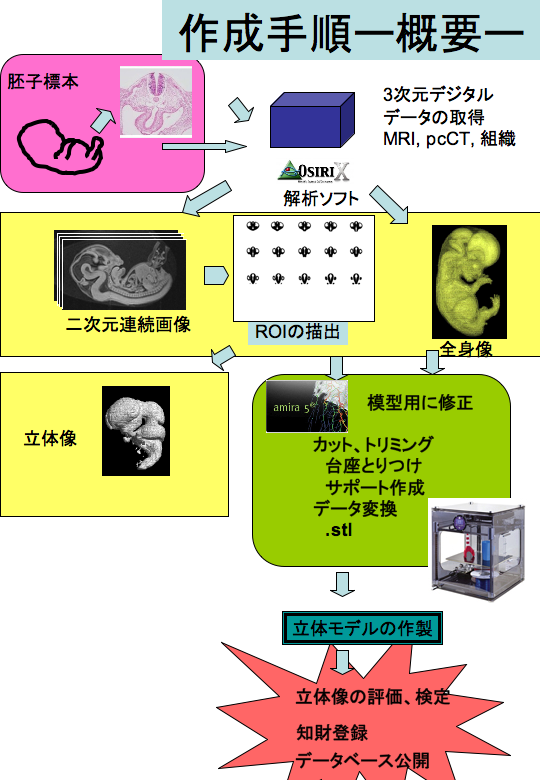
3Dプリンタとは、通常の紙に平面的に印刷するプリンタに対して、3D CAD、3D CGデータを元に立体を造形する立体プリンタのことです。従来の方法としては、コンピュータ上で作った3Dデータを設計図として、液状の樹脂に紫外線などの光を照射し、少しずつプラスチックを硬化させることで 立体物を作るのが一般的でした。これに対して25年程前から「積層造形」という方法が開発され、3Dプリンタ−が低価格で供給されるようになってきました。

「積層造形」では、1mm以下の薄い樹脂や石膏などの材料を少しずつ積み重ねて立体模型を作るという方法です。ひも状のものを作ってぐるぐると輪を作って積み重ねて行く手法は縄文土器や焼き物の手びねりと共通のものがあります。データさえあれば”金型なしで”3Dプリンタが模型を自動で作ってくれます。その手軽さから、工業分野から医療、歯科、玩具、アートまで広く応用されつつあります。最近では、10万円前後の商品もあり、家庭でも購入できる価格帯になってきています。私たちは、MRIdataから3Dプリンタを用いて立体像の作成を行っています。

コンピュターグラフィックスは進歩し、display上で立体的にものをみることが可能になっていますが、実際にモデルを手に取ると、イメージと随分違ったり、新たな発見があったりします。3Dプリンタを用いた立体模型は、ヒト胚子の研究や教育への応用が期待できると考えられます。昔Zieglerらのモデルがヒト発生を体現したように、3Dプリンタを用いた立体模型は新たな伝道師となりうるのでしょうか。

4. Shiraishi N, Yamada S, Takakuwa T. Three-dimensional Models Once Again – for research and teaching of early human development, Congenit Anom (Kyoto), 2013; 53(1), 58-59, doi: 10.1002/ar.22662

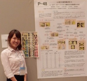
白石君が優秀ポスター賞を受賞(52th日本先天異常学会)

第52回日本先天異常学会で発表しました。(H24.7/6-8, 東京)

白石君の演題[3次元プリンタを用いたヒト胚子脳神経模型の作製]が優秀ポスター賞を受賞しました。内容とともに模型をポスターに直接貼付けるというインパクトが評価されたようです。
発表演題

ヒト胚子を用いた脳室形態形成と側脳室脈絡叢の解析 白石直樹

3次元プリンタを用いたヒト胚子脳神経模型の作製 白石直樹

Shiraishi N, Yamada S, Takakuwa T. Three-dimensional Models Once Again – for research and teaching of early human development, congenital anomaly, in press

ヒト胚子の外耳の動きは”分化・成長”で説明できる 高桑徹也、神楽所みほ

神楽所さんの卒業研究です。Head Face Med. 2012 Feb 1;8(1):2. (概要)

肝臓に形態形成異常があるヒト胚子のスクリーニング 金橋 徹

田中さんの卒業研究を継続、発展させています。