


岩佐さんの僧帽筋形成についての論文がJ Anatomyに受諾されました。MRI_DTIを用いて僧帽筋形成過程を、外観だけでなく、椎体レベルごとの筋線維の走行まで観察しました。
- 胎児初期におけるヒト僧帽筋(TpzM)の発達を観察し、拡散テンソル画像(DTI)分析を適用して生理機能につながる筋肉の構造を記述
- 僧帽筋はCS20で初めて検出され、その位置は、その挿入部および起始部である肩甲骨、鎖骨、および椎骨の形成とともに変化した。
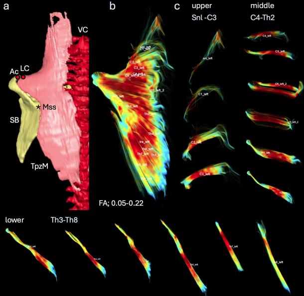
- 腹側ビューでの線維配向は、挿入部が変化する中間部分を除いて、頸椎から胸椎にかけて徐々に変化し、これはすべての初期胎児標本でほぼ同じであった。
- 僧帽筋の上部では、椎骨レベルに応じて線維配向が3次元的になだらかに変化していた。
69. Iwasa Y, Kanahashi T, Imai H, Otani H, Yamada S, Takakuwa T. Human trapezius muscle development during early fetal period. J Anatomy 2024, in press, doi: 10.1111/joa.14116
Abstract
This study aimed to observe human trapezius muscle (TpzM) development during the early fetal period and apply diffusion tensor imaging (DTI) analysis to describe the muscle architecture that leads to physiological functions. Human embryonic and early fetal specimens were selected for this study. TpzM was first detected at Carnegie stage 20. The position of the TpzM changed with the formation of the scapula, clavicle, and vertebrae, which are its insertions and origins. DTI revealed the fiber orientation from each vertebral level to dissect each muscle. Fiber orientation in the ventral view gradually changed from the cervical to thoracic vertebrae, except for the middle part at which the insertions changed, which was almost similar in all early fetal specimens. The TpzM volume increased from C1 to C7 in the upper part, reached local maxima at C6 and C7 in the middle, and then decreased. These muscles can be categorized into three parts according to their insertions and presented with the features of each part. The fiber orientation and distribution of the three parts at the vertebral level were almost constant during the early fetal period. The border between the upper and middle parts was mainly located around the C6 and C7 vertebral levels, whereas the middle and lower parts were between the Th1 and Th2 vertebral levels. A three-dimensional change in the fiber orientation in the upper part of the TpzM according to the vertebral level was noticeable. Our data will help to elucidate the developmental processes of TpzM.