磯谷さんの修士論文がAnatomical Recordに掲載されました。
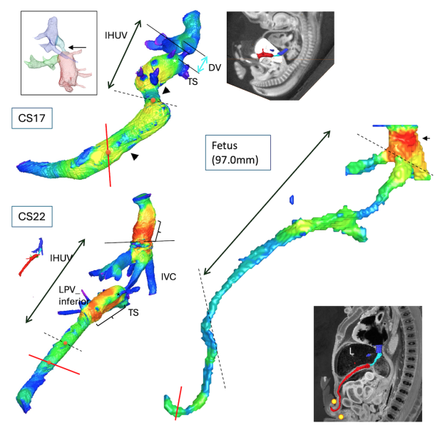
胎児循環に特有の胎盤から心臓にむかう静脈路(臍帯静脈、門脈洞、静脈管、下大静脈)について、領域による特徴を高解像度デジタルデータ(MRI, CT)から得られた立体再構成像と組織像を用いて検討しました。
- 胎児循環に特有の胎盤から心臓にむかう静脈路(臍帯静脈、門脈洞、静脈管、下大静脈)について領域ごとの特徴を検討
- 胚子期から胎児期初期の29個の標本を高解像度デジタル画像化のために選択し、18個の胚を組織学的解析のために選択した。
- 領域による特徴を高解像度デジタルデータ(MRI, CT)から得られた立体再構成像と組織像を用いて検討し明らかにした。
67. Isotani N, Kanahashi T, Imai H, Yoneyama A, Yamada S, Takakuwa T. Regional differences in the umbilical vein and ductus venosus at different stages of normal human development. Anat Rec (Hoboken), 2024, 307, 3306-3326.DOI:10.1002/ar.25421
During the fetal period, oxygenated blood from the placenta flows through the umbilical vein (UV), portal sinus, ductus venosus (DV), and inferior vena cava (IVC) to the heart. This venous route varies regionally in many aspects. Herein, we sought to characterize the venous route’s morphological features and regional differences during embryonic and early-fetal periods. Twenty-nine specimens were selected for high-resolution digitized imaging; 18 embryos were chosen for histological analysis. The venous route showed a primitive, large, S-shaped curved morphology with regional narrowing and dilation at Carnegie stage (CS) 15. Regional differences in vessel-wall differentiation became apparent from approximately CS20. The vessel wall was poorly developed in most DV parts; local vessel-wall thickness at the inlet was first detected at CS20. The lumen of the venous route changed from a non-uniform shape to a relatively round and uniform morphology after CS21. During the early-fetal period, two large bends were observed around the passage of the umbilical ring and at the inlet of the liver. The length ratio of the extrahepatic UV to the total venous route increased. The sectional area gradually increased during embryonic development, whereas differences in sectional area between the DV, UV, and IVC became more pronounced in the early-fetal period. Furthermore, differences in the sectional area between the narrowest part of the DV and other hepatic veins and the transverse sinus became more pronounced. In summary, the present study described morphological, morphometric, and histological changes in the venous route throughout embryonic and early-fetal development, clarifying regional characteristics.