


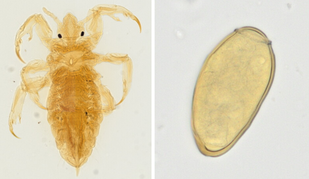
医動物学実習で使用する寄生虫スライド標本のデジタル化の取り組みについて発表しました。第36回日本臨床微生物学会総会(名古屋:2025/1/24-26)
バーチャルスライドを活用した寄生虫学・医動物学教育の試み:
金橋 徹、伊吹謙太郎、山田 稔、高桑徹也
|
||||
  |
医動物学実習で使用する寄生虫スライド標本のデジタル化の取り組みについて発表しました。第36回日本臨床微生物学会総会(名古屋:2025/1/24-26) バーチャルスライドを活用した寄生虫学・医動物学教育の試み: 金橋 徹、伊吹謙太郎、山田 稔、高桑徹也 卒業研究発表会が行われました (2025.1.27-28, 高井ホール) ヒト胚子期・胎児期における前腕骨の形態形成の解析 伊澤駿希 ヒト胚子期・胎児期における上腕骨の形態形成の検討 H.T. ヒト胚子期・胎児期初期に胎盤に流入する動脈系の解析 中井泰千 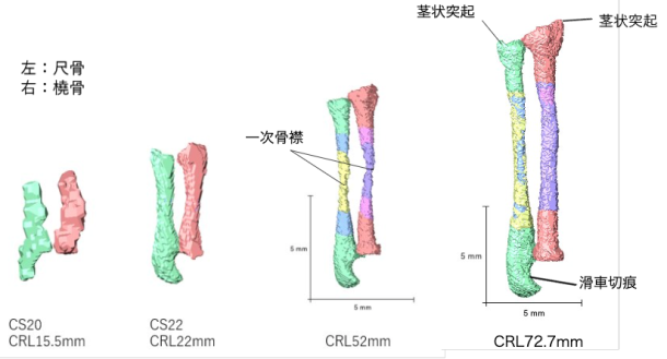 石田さんの修士論文の一部がJ Anatomyに受諾されました CS16からCS23までの合計47個のヒト胚の高解像度の磁気共鳴画像取得し、中腸のヘルニア期に中腸と腸間膜で時間の経過とともに生じる形態学的変化を検討、二次ループと三次ループの初期形成、それに続く追加ループの形成を正確に記載しました。この研究の知見は、ループ形成における遺伝的要因と生体力学的要因の役割を包括的に理解する上で極めて重要です。 71. Tanima M, Ishida N, Ueda Y, Kanahashi T, Matsubayashi J, Imai H, Yamada S, Takakuwa T.Hierarchical loop formation in human midgut during physiological umbilical herniation, J Anatomy 2024, in press DOI:10.1111/joa.14228 
Abstract The primary loop, a single hairpin-shaped loop, becomes recognizable at the Carnegie stage (CS) 16. This loop projects toward the umbilical cord and subsequently gives rise to four secondary loops in the midgut of human embryos. As development advances, the segments corresponding to each secondary loop further develop into an increasing number of loops, referred to as tertiary loops. The mesenteric leaves and the narrowing parts, which serve as the borders of the secondary loops, remain identifiable throughout the subsequent stages of development. This study aimed to describe the morphological alterations that occur in the midgut and mesentery over time during the herniated phase of the midgut. A total of 47 human embryos between CS16 and CS23 and two fetuses in the physiological umbilical herniated stage were selected for high-resolution magnetic resonance imaging acquisition. Specimens were obtained from the Congenital Anomaly Research Center of Kyoto University. Serial tissue sections obtained from four embryos were subjected to histological observation. The midgut and mesentery were reconstructed in three dimensions, and the resulting morphological changes were observed and analyzed. Formation of the primary loop was observed in all specimens between CS16 and CS18. Secondary loops in the midgut were initially discerned at CS19 in segments 2 and 4 (S2 and S4). The border between S3 and S4 was identified at the apex of the midgut hernia, where traces of the vitelline artery and duct enter the mesentery. At CS21 and later stages of development, the presence of three borders at the exact location delineated by mesenteric narrowing was consistently observed, which resulted in the midgut being divided into four segments in all specimens. The formation of tertiary loops was initially identified at Carnegie stage (CS) 21, occurring in either segment S2 or S3. By CS23, tertiary loops were observed in three segments in most specimens. Notably, the initial formation of tertiary loops in S4 occurred one Carnegie stage later than in S2 or S3. Additionally, the increase in the number of folds and the length per fold in S4 was delayed compared with the number and length of folds observed in both S2 and S3. The number of loops in S1 remained constant (one secondary loop) across all specimens. Upon reaching a critical threshold length, the number of loops exhibited a marked increase, accompanied by rapid elongation in S2, S3, and S4. The number of tertiary loops increased in accordance with the crown-rump length, which exhibited a maximum of 19 tertiary loops in S2 to S4 of the midgut. These findings support the hypothesis that tertiary loops develop biomechanically through the rapid elongation of the midgut and slow growth of the mesentery. This study describes the morphological alterations occurring in the midgut and mesentery over time during the herniated phase of the midgut and provides a comprehensive understanding of the roles of genetic and biomechanical factors in loop formation. 30. Three-dimensional structure of the human midgut with mesentery and factors determining midgut loop formation  【第一章:背景】胚子期・胎児期の中腸は、臍帯内で腸管ループを増加させた後、腹腔内 へ還納する。一連の研究は、経時的な腸管の形態学的変化の詳細な描出、および腸管ルー プの形成に影響を与える要因の検討を目的とした。 29. Three-dimensional analysis of the human embryonic and early fetal lens 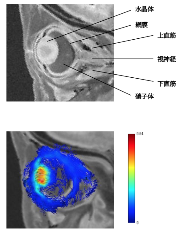 【背景】水晶体の発生はCarnegie stage(CS)13で眼胞が表皮外胚葉と接した後、表皮外胚葉が厚みを増して水晶体プラコードを形成することで開始する。CS15までに水晶体プラコードは表皮外胚葉と分離し、基底膜で囲まれた水晶体胞が形成される。水晶体胞の前壁は水晶体上皮になり、後壁の細胞はCS16以降に分化して前後方向に一次水晶体線維細胞を形成し、その後新しい線維細胞である二次水晶体線維細胞が一次水晶体線維の外側に追加され始める。この二次水晶体線維細胞は胎児期以降、出生後の成人期にも追加され続けるが詳しい伸長方向や、胎児期以降の水晶体の形態変化については明らかになっていない。今回、水晶体線維細胞の配向性を解明するために、拡散テンソル画像(DTI)を用いて解析を行った。DTIは水分子の拡散異方性の可視化が可能であり、T1強調画像では確認することのできない線維細胞の配向性の観察が期待される。 28. Development of the tongue in the human embryonic and fetal period 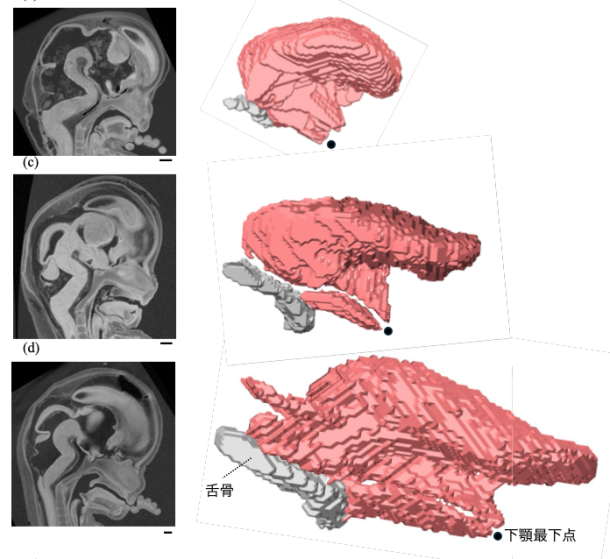 【背景】舌は8つの筋肉が複雑に交差した構造であり、舌筋の形成過程を把握するには三次元的な解析が必要である。また舌形成の観点から頭蓋顎顔面の形成過程を定量的に検討した例はない。 27. Three-dimensional analysis of human fetal tarsal development 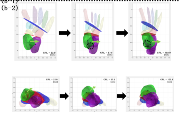 [方法]本研究では、ヒト胚子および胎児の固定標本 31 例を分析対象とした。MR 画像から 右足の足根骨・中足骨・脛骨・腓骨を立体化し、ランドマークを設定して 3 次元座標系を 構築した。この座標系を基に以下の解析を行った。1関節面角度の定量化。Chopart 関節 (横足根関節)、Lisfranc 関節(足根中足関節)、および Talocalcaneal 関節(距骨下関節) の関節面を定義し、主成分分析を用いて関節面の長径方向(第一主成分)および法線方向 (第三主成分)を算出した。これらのベクトルの角度を座標軸に基づき計測した。2足ア ーチ指標の定量化。内側縦アーチの評価を行うため、第一中足骨接地点・踵骨接地点を通 り、足底面と垂直な断面における Chopart 関節高、Lisfranc 関節高、足アーチの長さ、足 下部の面積を算出した。また、足下部に複数の測定点を設定し、その地面からの高さを足 アーチの指標として算出した。3足根骨の成長のシミュレーション。立体像の表面から複 数の点をサンプリングし、CRL を説明変数、各サンプリング点の座標を目的変数として 3 次回帰を行った。これにより、成長段階(CRL の増加)に応じた足根骨・中足骨の形状 の変化を予測した。 [結果]1関節面角度の定量化。Chopart関節面は、lateral viewではつま先側に倒れるよう に変化し、superior view では反時計回りに、posterior view では内側が持ち上がり時計回り に変化した。一方、Lisfranc 関節面も同様の傾向を示したが、その変化量は Chopart 関節 面よりも小さかった。Talocalcaneal 関節面は lateral view において、足底に対し傾いた状態 から、足底と水平になるまで変化した。また、lateral view における Talocalcaneal 関節面と Chopart 関節面の角度変化には強い相関が認められた。2足アーチ指標の定量化。Lisfranc 関節高と CRL との相関は弱かったが、Chopart 関節高、足下部の面積、足アーチの長さは CRL とやや強い相関を示した。3足根骨の成長のシミュレーション。CRL が大きくなる につれ、足根骨間の隙間が縮小し、骨同士が密集する傾向が確認された。また、距骨と踵 骨の位置関係や舟状骨の方向、中足骨群の長さに変化が見られた。 [結論]本研究では胎児の足根骨を 3 次元的に解析し、関節面角度の変化、足アーチ指標の 計測、成長シミュレーションを実施することで、不足している胎児における足根骨の成長 についてのデータを提供した。将来的には、胎児期における足部の発達メカニズムの解明 や、発達異常の早期診断への貢献が期待される。 |
|||