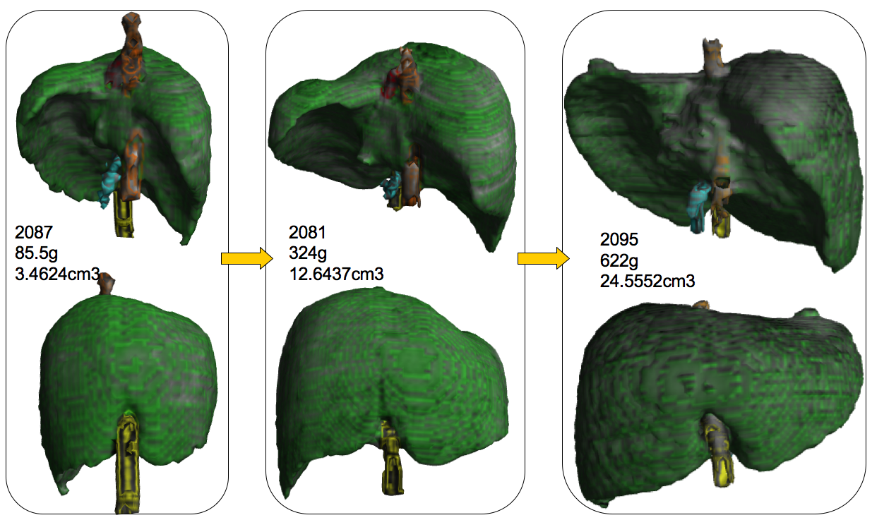
濱部さんの卒論がHepatol Resに掲載されました
- 妊娠中期(16〜28週)の肝臓形成の過程を検討
- 胎児期の肝臓は体重の増加に伴って横径・縦径・厚みすべてが増大
- 縦径に比べ、横径、厚みの増加が顕著
- 体幹部の成長に伴って肝臓も同じ比率で成長
- 分葉の割合はほぼ一定
5.Hamabe Y, Hirose A, Yamada S, Takakuwa T et al, Morphology and Morphometry of Fetal Liver at 16–26 Weeks of Gestation by Magnetic Resonance Imaging – Comparison with Embryonic Liver at Carnegie Stage 23, Hepatol Res,2013; 43: 639–647, doi: 10.1111/hepr.12000
Abstract
Aim
Normal liver growth was described morphologically and morphometrically using magnetic resonance imaging (MRI) data of human fetuses, and compared with embryonic liver to establish a normal reference chart for clinical use.
Methods
MRI images from 21 fetuses at 16–26 weeks of gestation and eight embryos at Carnegie stage (CS)23 were investigated in the present study. Using the image data, the morphology of the liver as well as its adjacent organs was extracted and reconstructed three-dimensionally. Morphometry of fetal liver growth was performed using simple regression analysis.
Results
The fundamental morphology was similar in all cases of the fetal livers examined. The liver tended to grow along the transversal axis. The four lobes were clearly recognizable in the fetal liver but not in the embryonic liver. The length of the liver along the three axes, liver volume and four lobes correlated with the bodyweight (BW). The morphogenesis of the fetal liver on the dorsal and caudal sides was affected by the growth of the abdominal organs, such as the stomach, duodenum and spleen, and retroperitoneal organs, such as the right adrenal gland and right kidney. The main blood vessels such as inferior vena cava, portal vein and umbilical vein made a groove on the surface of the liver. Morphology of the fetal liver was different from that of the embryonic liver at CS23.
Conclusion
The present data will be useful for evaluating the development of the fetal liver and the adjacent organs that affect its morphology.


