熊野くんの卒業研究のうち上肢の解析分がAnatomical Recに掲載されました。
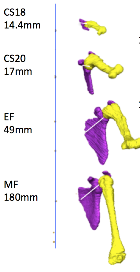
胚子期後期から胎児期初期、中期についての上肢の肢位について、上腕骨と体幹、体幹と肩甲骨、肩甲骨と上腕と解剖学的にわけて検討しています。
- 上腕の外転と屈曲ともに、CS20 で極大値を示し、外転は中期胎児期まで徐々に減少した。
- 肩甲骨は、関節腔が頭側および腹側を向いた独特の位置を示した。
- この独特な肩甲骨の位置は、肩甲骨_上腕関節の角度の鏡像的な変化により上腕の姿勢にほとんど影響を与えない。
Kumano Y, Tanaka S, Sakamoto R, Kanahashi T, Imai H, Yoneyama A, Yamada S, Takakuwa T. Upper arm posture during human embryonic and fetal development. Anatomical Rec 2022, 305 1682-1691, https://doi.org/10.1002/ar.24796
Abstract
The upper extremity posture is characteristic of each Carnegie stage (CS), particularly between CS18 and CS23. Morphogenesis of the shoulder joint complex largely contributes to posture, although the exact position of the shoulder joints has not been described. In the present study, the position of the upper arm was first quantitatively measured, and the contribution of the position of the shoulder girdle, including the scapula and glenohumeral (GH) joint, was then evaluated. Twenty-nine human fetal specimens from the Kyoto Collection were used in this study. The morphogenesis and three-dimensional position of the shoulder girdle and humerus were analyzed using phase-contrast X-ray computed tomography and magnetic resonance imaging. Both abduction and flexion of the upper arm displayed a local maximum at CS20. Abduction gradually decreased until the middle fetal period, which was a prominent feature. Flexion was less than 90° at the local maximum, which was discrepant between appearance and measurement value in our study. The scapular body exhibited a unique position, being oriented internally and in the upward direction, with the glenoid cavity oriented cranially and ventrally. However, this unique scapular position had little effect on the upper arm posture because the angle of the scapula on the thorax was canceled as the angle of the GH joint had changed to a mirror image of that angle. Our present study suggested that measuring the angle of the scapula on the thorax and that of the GH joint using sonography leads to improved staging of the human embryo.


